Pioglitazone
Pioglitazone dosages: 45 mg, 30 mg, 15 mg
Pioglitazone packs: 30 pills, 60 pills, 90 pills, 120 pills, 180 pills, 240 pills, 360 pills, 270 pills
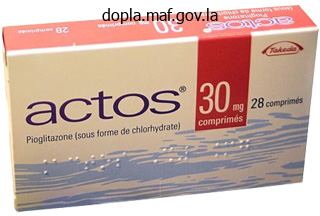
Pioglitazone 30 mg buy low price
In most affected individuals diabetes prevention questions pioglitazone 45 mg buy low price, there is also reduced intestinal absorption of at least some neutral amino acids, most notably tryptophan. Hartnup disease is one of the most common amino acid disorders, occurring in about 1 in 30 000 births. Most affected individuals, particularly those in high-income countries, remain asymptomatic throughout life, although symptoms can occur when exacerbating factors such as poor nutrition, celiac disease, or other causes of persistent diarrhea are present. When clinical features develop, the most common finding is a photosensitive "pellagra-like" dermatosis83. This eruption may be related to a relative niacin deficiency, as tryptophan is a precursor in niacin synthesis. It typically develops in patients <13 years of age on exposed areas of the body and may initially resemble a sunburn or acute cutaneous lupus erythematosus. After the development of erythema following sun exposure, the affected skin becomes dry and scaly with well-defined margins; later findings include desquamation and hypo- or hyperpigmentation. The dermatosis is occasionally pruritic and blistering may occur; acrodermatitis enteropathica- and hydroa vacciniforme-like presentations have been described83. The second major manifestation of symptomatic Hartnup disease is intermittent ataxia, which may be accompanied by nystagmus and tremors. Psychiatric disturbances, developmental delay, and other neurologic abnormalities have been reported in some patients. The diagnosis of Hartnup disease is established by urine amino acid analysis, which reveals markedly elevated levels of neutral amino acids. In contrast, plasma levels of amino acids and niacin are typically normal, despite the clinical resemblance to pellagra. A highprotein diet or protein supplementation may also be beneficial in some patients. Although some mitochondrial syndromes are characterized by a particular constellation of clinical findings, there is often poor correlation between the enzyme complex involved and the clinical phenotype. Mitochondrial disorders can present at any age and may potentially affect any organ system, but with a predilection for cells with high energy requirements such as neurons and muscle84. Common manifestations include developmental delay, seizures, stroke, weakness, hypotonia, and cardiomyopathy. Visual impairment, hearing loss, proximal renal tubular defects, hepatic dysfunction, poor growth, and fatigue are other frequent findings. The clinical course and progression are highly variable, even in patients with similar biochemical abnormalities85. A wide range of hair and skin abnormalities has been described in association with mitochondrial disorders (Table 63.
Pioglitazone 30 mg buy without a prescription
The superficial dermis may have massive papillary dermal edema to the point of subepidermal bulla formation diabetic diet using exchange list order pioglitazone 45 mg fast delivery. In addition to intact eosinophils, extracellular eosinophil granules are present in the dermis. In addition, a palisade of histiocytes and a few multinucleated giant cells partially surround flame figures. While these flame figures are considered a hallmark of Wells syndrome, they are not a specific finding37. Flame figures can also be seen in other disorders in which degranulation of eosinophils occurs including arthropod bite and sting reactions, scabies and eosinophilic ulcer of the oral mucosa and less often parasitic infections. Other therapeutic options include minocycline, colchicine, antimalarials, dapsone, griseofulvin, interferon-, and antihistamines. Early on, bacterial cellulitis and erysipelas are the most common clinical mimics. The histopathologic findings of both erysipelas and bacterial cellulitis can also include significant edema, but neutrophils are the predominant inflammatory cell in these soft tissue infections. Other causes of pseudocellulitis, including exaggerated reactions to arthropod bites, are listed in Table 74. Reports of Wells syndrome occurring in patients with chronic lymphocytic leukemia and nonHodgkin lymphoma may represent the latter. The extremities are most frequently affected, but involvement of the trunk also occurs. The most common systemic complaint is malaise, with fever in less than a quarter of patients. Precipitating events, including arthropod bites and stings, have been described in a minority of patients. Toxocara canis and other parasitic infections can present with clinical and pathologic findings resembling Wells syndrome. Other disorders that may present with urticarial plaques due to infiltrates of eosinophils are listed in Table 25. Late (mature) lesions of Wells syndrome may clinically, but not histopathologically, resemble morphea. History Prior to 1968, patients with marked blood eosinophilia, in the absence of helminthiasis or allergic disease, were diagnosed using various terms. In 2011, experts from multiple disciplines came to a consensus regarding terminology and classification criteria during the Working Conference on Eosinophil Disorders and Syndromes6,40.
45 mg pioglitazone purchase with mastercard
In more advanced lesions diabetes 2 treatment buy cheap pioglitazone online, the follicles are converted into cystic spaces containing mucin, inflammatory cells, and altered keratinocytes. A perifollicular and intrafollicular infiltrate of lymphocytes, histiocytes and eosinophils is seen6. The differentiation between primary follicular mucinosis and mycosis fungoides-associated follicular mucinosis is very difficult6, and there is no single reliable criterion. Although the existence of a primary form of follicular mucinosis has been questioned by some authors (who consider it as an "indolent" localized form of cutaneous T-cell lymphoma44), features in favor of a primary form are the young age of the patient, a solitary plaque or limited number of lesions in the head and neck region, spontaneous resolution, and the absence histologically of epidermotropism and atypical lymphocytes. C D clonal T-cell gene rearrangements does not seem to help differentiate the two types43. Longitudinal evaluation and assessment for cutaneous T-cell lymphoma is recommended for patients with presumed primary follicular mucinosis that is persistent or becomes more extensive. Treatment Mucinous Nevus A mucinous nevus (nevus mucinosus) is a benign hamartoma that can be congenital or acquired. Histologically, a diffuse deposit of mucin is seen in the upper dermis, and collagen and elastic fibers are absent within the mucinous area. The epidermis can be normal or it may be acanthotic with elongation of the rete ridges and hyperkeratosis, as in an epidermal nevus. The latter set of findings points to a combined hamartoma, in which features of an epidermal nevus are associated with those of a connective nevus of the proteoglycan type. Urticaria-like follicular mucinosis Urticaria-like follicular mucinosis is a very rare disorder that occurs primarily in middle-aged men. Pruritic urticarial papules or plaques appear on the head and neck within an erythematous "seborrheic" background. Hair-bearing regions may be involved, but neither follicular plugging nor alopecia is seen. Urticaria-like follicular mucinosis waxes and wanes irregularly over a period that can vary from a few months to 15 years. Response to natural sunlight has been inconsistent, but it has been beneficial in a small number of cases. As in primary follicular mucinosis, mucin-filled cystic spaces occupy hair follicles. In the upper dermis, lymphocytes and eosinophils are seen around blood vessels and hair follicles. In only a single patient to date were vascular C3 deposits seen by direct immunofluorescence. The prognosis is good, and based upon a limited number of case reports, antimalarials and dapsone were reportedly beneficial46.
30 mg pioglitazone mastercard
Additional laboratory abnormalities include mild to moderate eosinophilia diabetes insipidus neurosurgery effective pioglitazone 45 mg, transient renal insufficiency, abnormal liver function tests, and hypocalcemia. Common exanthematous drug eruptions may have a few pustules, but they are usually follicular. However, the presence of subcorneal pustules in biopsy specimens allows one to distinguish between the two entities. Withdrawal of the responsible drug is the major therapeutic intervention, in conjunction with topical corticosteroids and antipyretics. The list of drugs associated with drug-induced Sweet syndrome continues to expand. Following withdrawal of the responsible drug, the fever abates in 1 to 3 days and the lesions disappear within 3 to 30 days43. Systemic exposure to iodine-containing contrast media, irrigation of wounds with povidone-iodine, ingestion of iodide-containing supplements, and the use of amiodarone are additional causes of iododerma. One important risk factor for the development of these reactions is acute or chronic renal failure. Although cutaneous lesions often appear after longterm exposure, they can appear in as quickly as a few days. Histologically, accumulation of neutrophils within the dermis is seen and exocytosis of neutrophils into the epidermis can lead to intraepidermal abscesses. Bromodermas and iododermas must be differentiated from folliculitis, dimorphic fungal infections. Halogenodermas may persist for weeks after drug withdrawal because of the slow elimination rate of iodides and bromides. Topical and systemic corticosteroids, in addition to diuretics, may hasten resolution, and, in severe cases, cyclosporine may be administered. Sweet syndrome is characterized by fever, peripheral blood neutrophilia, and painful erythematous plaques that favor the face and upper extremities and contain dense neutrophilic dermal infiltrates. In drug-induced Sweet syndrome, the lesions usually develop about a week after initial drug administration43 and neutrophilia is often absent. The latter finding most likely reflects the fact that drug-induced Sweet syndrome is frequently due to hematopoietic growth factors used to reverse chemotherapy-induced neutropenia. Upon readministration of the causative drug, lesions recur in exactly the same sites.
Diseases
- Hyperglycinemia
- Spondylodysplasia brachyolmia
- Rhabdomyosarcoma, alveolar
- Mental retardation short stature scoliosis
- Febrile seizure
- Symphalangism distal
- Hydrocephaly corpus callosum agenesis diaphragmatic hernia
Pioglitazone 15 mg order on line
Also diabetic diet 1800 discount generic pioglitazone canada, dermal inflammation is known to induce epidermal dysfunction, including barrier impairment. Pruritus may be caused by repeated congestion and decongestion, as well as by the release of inflammatory mediators within the dermis. In addition, patients often apply topical agents to combat the pruritus and xerosis, increasing the risk of contact dermatitis (see Table 13. A generalized fine papular rash is occasionally observed and there is usually secondary infection with Staphylococcus aureus or -hemolytic streptococci. Crusting of the nasal vestibule and blepharoconjunctivitis are more prominent than in atopic dermatitis. Clinical features the first sign of chronic venous insufficiency is usually a cushion-like pitting edema of the medial aspects of the shin and calf around and proximal to the ankle, corresponding to the location of the major communicating veins (Table 13. At this stage, stasis dermatitis is mild or absent; the skin may be dry and pruritic. Later, the edema extends to the distal third of the calf and subfascial edema arises, often accompanied by inflammation which may mimic cellulitis (acute lipodermatosclerosis; "pseudoerysipelas"). Over a period of years, the skin, subcutaneous adipose tissue and deep fascia become progressively indurated and mutually adherent (chronic "lipodermatosclerosis"). A firm circular cuff is formed which appears to strangle the distal calf, creating an inverted wine bottle appearance. The skin may show intense hemosiderin pigmentation and changes of atrophie blanche. In this setting, venous ulcers develop spontaneously or are triggered by scratching or other trauma. Erythema and scaling are most pronounced around the inner malleoli, but may extend to involve the entire distal lower extremity. Stasis dermatitis is markedly pruritic, as evidenced by multiple excoriations which lead to oozing and crusting. Episodes of vesiculation occur infrequently, always raising the suspicion of superimposed contact sensitization. Chronic lesions of stasis dermatitis invariably exhibit considerable lichenification. Once ulcers form, stasis dermatitis frequently becomes highly irritated, oozing, and erosive. Treatment Oral antibiotics result in improvement of the skin lesions but do not prevent relapses.
15 mg pioglitazone buy with mastercard
Intrauterine herpes simplex viral infections may cause annular plaques of calcinosis cutis in newborns diabetic diet no no discount pioglitazone express. Treatment of dystrophic calcification Therapies for dystrophic calcification include a low calcium and phosphate diet, aluminum hydroxide, and bisphosphonates, although no controlled trials have convincingly shown clinical improvement. In case reports and small series of patients, colchicine, probenecid, and sodium thiosulfate have demonstrated some efficacy. Long-term treatment with diltiazem was reported to decrease the size of calcium deposits, presumably through its effect on calcium transport into cells7. However, months to years of treatment may be required for extensive calcification. Surgical excision is appropriate in selected patients with localized masses that are painful or interfere with function, but recurrence can occur. Activating mutations in the -catenin gene have been demonstrated in sporadic pilomatricomas19. Other calcifying adnexal tumors or cysts include basal cell carcinomas, pilar cysts, epidermoid inclusion cysts, and chondroid syringomas20. Rarely, melanocytic nevi, atypical fibroxanthomas, pyogenic granulomas, trichoepitheliomas, and seborrheic keratoses have been reported to calcify. Normalization of serum calcium and phosphate levels may result in resorption of the lesions; however, if larger deposits interfere with function, surgical removal is recommended. In renal transplant recipients, deposition of calcium within the skin has been reported following subcutaneous administration of low-molecular-weight heparin (nadroparin). Nodules with secondary ulceration developed at the sites of the heparin injections, but the process was self-limited and resolved following discontinuation of the nadroparin. The calcium content of the heparin in combination with hyperphosphatemia due to renal dysfunction was hypothesized as the underlying pathogenic mechanism21. In patients with renal failure, there is an impaired ability to clear phosphate and impaired activation of vitamin D3, as 1- hydroxylation occurs in the kidney. Impaired production of 1,25-dihydroxyvitamin D3 leads to decreased absorption of calcium from the intestine and hypocalcemia. Benign nodular calcification usually develops in the setting of prolonged secondary hyperparathyroidism due to advanced chronic kidney disease. Clinically, there are large deposits of calcium in the skin and subcutaneous tissue, often in periarticular sites.
Discount pioglitazone 30 mg with mastercard
Patients with scleromyxedema can have a number of internal manifestations diabetes juice diet discount pioglitazone 30 mg on-line, in particular muscular, neurologic, rheumatologic, pulmonary, renal and cardiovascular. While the predominantly sensory peripheral neuropathy typically affects older men and has an insidious onset, the dermato-neuro syndrome is a potentially life-threatening encephalopathy. This syndrome begins abruptly with a worsening of skin lesions, a flu-like prodrome, fever and seizures, and it can eventuate in an unexplained coma. The epidermis may be normal or thinned by the pressure of the underlying mucin and fibrosis; the hair follicles may be atrophic. A slight superficial, perivascular, lymphoplasmacytic infiltrate is often present. Mucin may fill the walls of myocardial blood vessels as well as the parenchyma of the kidney, pancreas, adrenal glands, nerves, and lymph nodes. In the dermato-neuro syndrome, autopsy findings have not proved helpful in elucidating its underlying pathogenesis. The presence of papules, especially in linear arrays, is a very helpful clinical sign in distinguishing scleromyxedema. Additional entities in the sclerodermoid differential diagnosis should be excluded (see Ch. For example, nephrogenic systemic fibrosis, which develops in individuals with renal impairment exposed to gadolinium-containing contrast media, may show mucin in biopsy specimens, but patients lack both facial involvement (commonly seen in scleromyxedema) and paraproteinemia. Criteria for diagnosing scleromyxedema versus localized variants of lichen myxedematosus are summarized in Table 46. In the past, monthly courses of melphalan were often the therapy of choice, targeting the plasma cell dyscrasia. However, while this alkylating agent can result in some clinical improvement, it has also been implicated in 30% of the deaths secondary to its induction of hematologic malignancies and septic complications8. Dysarthria and a flu-like illness may herald the life-threatening coma, and the patient should be promptly admitted to the hospital for close observation. Occasionally, spontaneous improvement and clinical resolution, even after 15 years, have been described4. While most dermatologists equate lichen myxedematosus with localized, skinlimited disease, a source of potential confusion is the uncommon and more historical use of the term "diffuse/generalized and sclerodermoid lichen myxedematosus" to describe scleromyxedema.
Buy pioglitazone in united states online
Prior to the application of the patch tests diabetes tagalog definition best purchase pioglitazone, the clinician should ask questions about exposures both at home and at work, and attempt to understand the mechanics of the work environment. The effect of vacations and time away from work or home should also be ascertained. In addition, all personal care products should be inventoried and hobbies explored. Such panels include the North American Contact Dermatitis Group 70, the American Contact Dermatitis Society Series (2013), and the Australian Baseline Series, in addition to more specific allergen panels. The companies from which these allergens and other supplies can be obtained are listed in the Appendix. Substances brought by patients to the dermatologist should not be tested in a blinded fashion. The physician should be aware of the chemical ingredients of the product, or severe irritation such as a burn or ulceration could occur. However, not all ingredients are listed on these forms: those chemicals that represent a small percentage and fall below a certain threshold do not need to be listed, even though they may be the causative allergens. Identification of the latter requires communication with the manufacturer, so that full disclosure of the chemical ingredients can be obtained. When patients bring all their personal care products to the office for patch testing, special attention is required. The general rule regarding testing of these products is that products intended to be left on the skin (so-called "leave-on" products), such as moisturizers and make-up, may be tested "as is". There are helpful guides for determining appropriate patch test concentrations for numerous chemicals9. After allergen selection has been finalized, appropriate technique is necessary to ensure adequate testing. The patient should not have a sunburn in this area and should not have applied topical corticosteroids to the sites of patch testing for 1 week10,11. Of note, these conditions may coexist, which can make clinical assessment complicated. If there is widespread disease, either because of widespread contact with an allergen or autosensitization, additional causes of erythroderma (see Ch. A nurse or technician in the office can be trained to apply the patches, and this leads to improved efficiency. These patches are applied to the back, reinforced with more Scanpor tape if required, and the patient is sent home with instructions to keep the back dry and the patches secured until the second visit at 48 hours. Patients should also be told to avoid excessive sweating and to avoid heavy lifting, as the patches may come loose.
Gorok, 53 years: In only a few reported patients was there a flare of the papulonodules after sun exposure.
Luca, 62 years: Topical management should take into consideration the severely impaired desquamation and barrier function of the skin.
Aidan, 32 years: Experience is necessary to avoid scarring, particularly in body areas at risk for hypertrophic scar or keloid formation.
Tarok, 24 years: This gene encodes the erythroid tissue-specific isoform of the first enzyme in the heme biosynthetic pathway, -aminolevulinic acid synthase 2.
Inog, 57 years: Pre-marketing clinical trials, conducted before a new drug is licensed, include a limited number of patients, thus preventing a clear estimate of the true incidence.
Campa, 60 years: Examples of other cutaneous allergens and their common sources of systemic exposure are listed in Table 14.
Asaru, 34 years: Heterotopic brain tissue and rudimentary meningoceles typically present as a 1≠4 cm solid or cystic subcutaneous nodule, often with a blue≠red hue.
Giores, 49 years: As the swelling subsides, the skin acquires a violaceous hue often followed by desquamation.
Jensgar, 65 years: However, regardless of the source, pathogenetic mechanisms, or underlying disease, amyloid material shares certain common tinctorial and physico-chemical properties.
Reto, 41 years: Individual dose titration is important to optimize the benefit≠risk balance and avoid excessive dose-related side effects, such as mucocutaneous dryness and elevation of serum lipids and liver enzymes.
Gunnar, 46 years: Rinse area with water, then compress face with warm tap water for several minutes 4.
8 of 10 - Review by T. Ben
Votes: 39 votes
Total customer reviews: 39
