
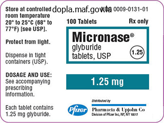
Micronase
Micronase dosages: 5 mg, 2.5 mg
Micronase packs: 90 pills, 120 pills, 180 pills, 360 pills
Buy 2.5 mg micronase with amex
The earliest signs of hypovolaemia are a rise in respiratory rate diabetes diet who micronase 2.5 mg without prescription, a delay in capillary refill time, and an abnormality of the cardiotocograph. Hypotension may be a late sign in young healthy women who compensate well initially. Women with these signs should be stabilised and immediately transferred to a consultant-led unit or any facility where an emergency delivery and transfusion of the mother can be performed. All units should have a massive obstetric haemorrhage protocol which includes contacting the haematologist on call for advice regarding the use of blood products. In women who are stable, the history should include the nature and amount of bleeding, the presence of pain, contractions, ruptured membranes, placental localisation at the last ultrasound scan, the result of the last cervical smear, and the rhesus status. Sudden onset of painless, bright red vaginal bleeding may suggest placenta praevia. Women with placenta praevia may have a few episodes of warning bleeds before a major bleed, and admission for observation needs to be considered. Sudden onset of a severe continuous abdominal pain associated with darker bleeding may suggest placental abruption. If the bleeding is associated with a thick mucoid plug, this may be a heavy show associated with the onset of labour. These vessels may rupture with either spontaneous or artificial rupture of membranes, causing significant fetal haemorrhage and even death. A soft, non-tender abdomen with a high unengaged presenting part or abnormal lie is suggestive of placenta praevia. The uterus may be markedly tender all over or localised to where the placental abruption has occurred. It can be due to multiple mild abruptions (separations) at the periphery of the placenta. It is important that the massive obstetric haemorrhage protocol is activated, appropriate blood products are used with haemotological advice, and active management of the third stage of labour and early detection and treatment of a consumptive coagulopathy are done to prevent this from happening. If the woman has not been booked in the index pregnancy, an ultrasound scan must precede a digital or a speculum vaginal examination to exclude a major placenta praevia, as examination may aggravate the bleeding. A speculum examination may help diagnose local causes of vaginal bleeding as well as help assess cervical dilatation. As mentioned earlier a transvaginal ultrasound is more accurate than a transabdominal ultrasound. Ultrasound may not be helpful in an early abruption where the diagnosis is usually a clinical one. This is by definition a retrospective diagnosis of amenorrhoea of 1-year duration due to failure of ovarian function. Any vaginal bleeding that occurs after 6 months of amenorrhoea from presumed menopause should be treated as suspicious. Investigations should be directed to determine the cause of bleeding depending on the age of the woman.
Buy micronase 2.5 mg with mastercard
Lymphoma Sometimes hepatic involvement in systemic lymphatic malignancy may result in anechoic masses in the liver diabetes prevention ideas cheap micronase 2.5 mg with visa. One typical feature is the simultaneous presence of the various stages of lymphatic infiltration: apart from solitary anechoic masses (no or slight posterior enhancement), there are often irregular areas of varying size with hypoechoic infiltration of the parenchyma. In addition, extrahepatic criteria such as splenomegaly with infiltrative changes and lymphadenopathy may be seen. Infiltrating Lymphoma Involvement of the liver is seen in about 50% of lymphoma cases (Hodgkin disease and non-Hodgkin lymphoma). Particularly the more advanced stages will demonstrate hepatic involvement with circumscribed malignant lymphadenopathy. Diffuse, miliary, or periportal infiltration of the parenchyma is primarily found in early stages of the disease and usually escapes ultrasound detection. In diffuse nonspecific parenchymal changes, the presence of splenic pathology and lymphadenopathy may warrant the presumed diagnosis of diffuse infiltrative lymphoma of the liver. Non-Hodgkin lymphoma with multiple hypoechoic masses and one almost completely anechoic lesion. Diffuse metastasis of the small nodular type may present as numerous anechoic masses that differ from polycystic liver disease by their irregular shape and diffuse margin. Vessels/Bile Ducts Any differential diagnosis of anechoic masses in the liver has to include the possibility of vessels or bile ducts being viewed in crosssection. In this case, the characteristic aspects are the tubular shape of the lesion when imaged in a cross-section plane, and its course, which can be traced all the way back to the originating vessel/duct. Bile ducts appearing as anechoic masses in the liver have to be pathologically enlarged/engorged. These cases are characterized by their periportal location and identification of the socalled hepatic triad, i. Being part of the hepatic triad, the branches of the portal vein course through the periportal area as well. Venectasia, varicosities, and collaterals in case of portal hypertension will all be imaged as enlarged segments of the intrahepatic tree of the portal venous system. They are easily identified by their typical course terminating at their stellate junction with the vena cava. The sonographer should keep in mind the possible venectasia and anomalies of the BuddChiari syndrome. These lesions may differ in size and distribution, but in most cases they are small and tend to have an irregular/diffuse margin. Although one cannot, in principle, deduce the type of tumor from the sonographic image alone, these hypoechoic metastatic deposits are quite frequent in undeveloped, undifferentiated, and actively growing malignancies. The finding of hypoechoic metastases is rather common (but not pathognomonic) in breast cancer, in small cell lung cancer, and in endocrine tumors.
Diseases
- Vasculitis hypersensitivity
- Dicarboxylicaminoaciduria
- Familial porencephaly
- Acromesomelic dysplasia, Maroteaux type
- Craniofaciocervical osteoglyphic dysplasia
- Brachytelephalangy characteristic facies Kallmann
- Cystathionine beta synthetase deficiency
- Bone marrow failure neurologic abnormalities
- Herpes simplex disease
- Heart situs anomaly
Micronase 5 mg with amex
These tumors have smooth margins blood sugar and stress micronase 2.5 mg visa, are found in the layered wall of the stomach, quite often relate to one of the central layers, and are well vascularized. Although these firm, elastic tumors display a somewhat varied echogenicity, they are mostly hypoechoic. Frequently, sonography will demonstrate a pathological concentric gut signature limited to the gastric antrum or cardia, while focal pathological atypical (off-center) gut signatures are more common in the corpus and rare in the fundus of the stomach. The typical lesion is visualized as a pathological thickening of the gastric wall with loss of the usual layering. Quite often the lesion is poorly delineated at the gastric lumen, and in case of concentric tumors it is not uncommon to encounter a stenotic lumen; in such cases there will be retention of food proximal to the stenosis. The immediate vicinity of the tumor shows a characteristic loss of peristalsis, while the pathological gut signature is hard and not compliant. Thickened, ovoid, pathological gut signature of the gastric antrum in the longitudinal plane; caudally, an enlarged pathologic lymph node. Lymphoma Gastric lymphoma may be visible on ultrasound as a focal hypoechoic mass involving the wall of the stomach, with clearly evident hypervascularity and loss of its typical layered appearance. At the gastric fundus and cardia, as well as at the distal esophagus, they may protrude transmurally into the lumen of the stomach as esophageal or fundic varices. Color flow Doppler scanning will demonstrate a ribbonlike hepatofugal portovenous blood flow. Pathomorphologically, it is a focal arteriovenous malformation, more precisely a vascular ectasia in the antrum of the stomach. Demonstration of a thick pathologic hypervascularity of the gastric antrum like an arteriovenous malformation. Ulceration Most gastric ulcers are far too small to be visualized on transabdominal ultrasound. At the ulceration, the gastric wall is thinned out and has lost its layered appearance; trapped air or food particles will often be seen within the cavity of the ulcer, resulting in a coarse echo. Diverticula, Mucosal Folds Diverticulum-like sacculation in the gastric wall is rare; when looking at all wall layers there will be focal excavation of the lumen. Transverse sections through rugal hypertrophy at the gastric body may mimic focal polypoid wall lesions. On ultrasonography, the pylorus appears as a circumscribed lengthy circular thickening of the muscle, the total pyloric diameter being in excess of 12 mm and the pyloric canal being compressed (so-called "cervix sign"); in addition, the stomach will display marked fluid retention. So-called cervix sign with marked circumscribed thickening of the lamina propria mucosae and tight pyloric canal, accompanied by gastric distension and fluid retention. Extended Wall Changes Gastrointestinal Tract Stomach Focal Wall Changes Extended Wall Changes Dilated Lumen Narrowed Lumen Small/Large Intestine Carcinoma/Scirrhus Lymphoma Gastritis Congestion, Edema Peritoneal Carcinomatosis Carcinoma/Scirrhus Adenocarcinoma of the stomach, especially scirrhous carcinoma, is characterized by diffuse extensive invasion of the gastric wall by the tumor, resulting in a narrowed lumen and loss of the layered wall architecture.
2.5 mg micronase purchase with amex
The significance of contrast enhancement in differentiating between benign polypoid masses and malignant tumors is still unknown blood sugar 10 day detox diet 5 mg micronase with mastercard. General Tumor Gallbladder Changes in Size Wall Changes General Hypoechogenicity General Hyperechogenicity Focal Hypoechogenicity/Hyperechogenicity General Tumor Focal Tumor Intraluminal Changes Nonvisualized Gallbladder Adenomyomatosis Cholesteatosis Gallbladder Carcinoma Adenomyomatosis Adenomyomatosis is a special case of gallbladder cholesteatosis and belongs to the group of hyperplastic cholecystoses. It appears as a hyperechoic pseudotumorous thickening of the wall (generalized or focal), originating from hypertrophied RokitanskyAschoff sinuses. Gallbladder carcinoma lacks the RokitanskyAschoff sinuses and intramural comet-tail artifacts. Focal Tumor Gallbladder Changes in Size Wall Changes General Hypoechogenicity General Hyperechogenicity Focal Hypoechogenicity/Hyperechogenicity General Tumor Focal Tumor Intraluminal Changes Nonvisualized Gallbladder Cholesterol Pseudopolyps Adenomas and Papillomas Adenomyomatosis Gallbladder Carcinoma Metastasis In order of frequency, focal changes in the gallbladder wall are mural polypoid lesions, partial wall thickening, and infiltrating lesions. They are hyperplastic polypoid neoplasms, usually hyperechoic, smoothly contoured, round or ovoid. Sometimes they are visualized as small hyperplastic (often hyperechoic) tumors, pendulant from the gallbladder wall, that may display a short stalk (hypoechoic lobulation is rather rare). Cholesterol polyps measuring more than 1 cm in diameter are the exception and therefore should undergo surgery. The detection of cholesterol pseudopolyps is highly dependent on the quality of the equipment. The most recent studies show that cholesterol pseudopolyps 1 mm in size can be detected (in up to 10% of all cases examined). Multiple small sessile hyperechoic polyps of 35 mm at the gallbladder wall (some with longer stalks). Usually, they are hypoechoic sessile structures resting on the intraluminal wall and are frequently found in the neck of the gallbladder. The differentiation of adenomas from papillomas relies on the fact that papillomas tend to be pedunculated, displaying a lobulated surface. All polyps are easily differentiated from calculi by their sessile growth, although sometimes viscous pseudopolypoid sludge may mimic a polyp. Combined with the focal cystic inclusions and the classic comet-tail artifacts, the focal hypertrophied AschoffRokitansky sinuses will round out the characteristic image in sonographic morphology. In segmental adenomyomatosis the gallbladder displays segmental constriction at the body. Gallbladder Carcinoma Every polypoid mass of irregular shape within the lumen of the gallbladder as well as every infiltrating lesion with destruction of the normal wall should be highly suspicious for gallbladder carcinoma. Gallstones and chronic cholecystitis play an important role in the carcinogenesis: 23% of people with gallstones will develop a carcinoma of the gallbladder. Most tumors appear hypovascular in color-flow duplex scanning, but a chaotic vascular pattern can be detected in suspected gallbladder carcinoma. The remaining gallbladder is without vessels delimiting the tumor from the gallbladder. Metastasis in the gallbladder wall is a rare finding and cannot be clearly differentiated by sonography from true gallbladder carcinoma. Intraluminal Changes Hyperechoic Gallbladder Changes in Size Wall Changes Intraluminal Changes Hyperechoic Hypoechoic Nonvisualized Gallbladder Gallstones Gallstones Gallstones are the classic example of mobile changes within the gallbladder.
2.5 mg micronase buy with visa
Convulsions blood glucose watch purchase 5 mg micronase with amex, hypertension crisis and acute renal failure in postpartum: role of bromocriptine? Emergency department use during the postpartum period: implications for current management of the puerperium. Delayed postpartum preeclampsia and eclampsia: demographics, clinical course, and complications. Postpartum toxemia: hypertension, edema, proteinuria and unresponsiveness in an unknown female. Perioperative hypertensive crisis and hemorrhagic diathesis: fatal complication of clinically unsuspected pheochromocytoma. Hemodynamics associated with the diagnosis and treatment of pheochromocytoma in pregnancy. Refractory hypotension during spinal anesthesia for Cesarean delivery due to undiagnosed pheochromocytoma. Outcomes of concurrent Caesarean delivery and pheochromocytoma resection in late pregnancy. Abnormal endothelium-dependent microvascular reactivity in recently preeclamptic women. Work, leisure-time physical activity, and risk of preeclampsia and gestational hypertension. Treatment of hypertension in pregnancy with methyldopa: blood pressure control and side effects. Antihypertensive treatment in pregnancy: analysis of different responses to oxprenolol and methyldopa. Fall in mean arterial pressure and fetal growth restriction in pregnancy hypertension: a meta-analysis. Fall in mean arterial pressure and fetal growth restriction in pregnancy hypertension: an 417 166. Maternal hypertension with superimposed pre-eclampsia: effects on child development at 71/2 years. The safety of calcium channel blockers during pregnancy: a prospective, multicenter, observational study. A plethora of national guidelines offer contrasting views regarding the hypertensive level at which drug therapy should be given during pregnancy and the authors in the initial sections of the chapter succinctly chronicle the reasons for this confusion. Additionally, systematic reviews of trials are discussed that further explain dilemmas in interpretation and provide guidelines for better-designed trials.
Syndromes
- Muscle twitching
- Right-lower quadrant
- Paralysis
- Growth hormone (GH) -- stimulates growth of tissues and bone
- Warm the person. If necessary, use your own body heat to aid the warming. Apply warm compresses to the neck, chest wall, and groin. If the person is alert and can easily swallow, give warm, sweetened, nonalcoholic fluids to aid the warming.
- If it leaks through to a part of the intestines, it is called an entero-enteral fistula.
- Headache with fever
- You will usually be asked not to drink or eat anything for 8 hours before the surgery.
- Sertofren
- ECG
Cheap 5 mg micronase free shipping
In cases of suspected widespread abdominal malignancies metabolic disease liver purchase micronase with amex, additional tumour markers should be used to investigate the possible primary. The causes for these situations can be divided into two main groups: neurogenic and non-neurogenic. In general, the anatomical level of the neurological injury determines the functional effect on the urinary system (Box 2). Spinal lesions may result in dyssynergia, an uncoordinated muscle contraction (see detrusor sphincter dyssynergia above), whereas sacral, cauda equina, or other peripheral nerve injuries may produce detrusor areflexia or absence of reflex. However, the situation is often more complex than this, as multiple or incomplete injuries can produce mixed patterns. Consequently, the functional result of the injury can often only be determined by video cystometrogram. This has the added bonus of allowing the measurement of the estimated pressure generated by the detrusor muscle during filling and micturition phases. This is important because, if the urinary system is exposed to an abnormally high functioning pressure, Further reading Green-top Guideline 62: Management of suspected ovarian masses in premenopausal women. Classic examples of this include avoiding faecal impaction and overdistension after surgery. An obstructed urinary system that is infected should be treated as an emergency, as septicaemia can result. If the obstructed lower urinary tract has resulted in renal dysfunction, an exact measurement of fluid input and urine output is undertaken to exclude a post-obstructive renal diuresis. If the urine output is excessive after catheterisation (>200 mL/h), intravenous fluids (normal saline) should be commenced to stop dehydration from occurring by initially balancing fluid input against the measured output. In the majority of patients with normal renal function and no urinary infection present, clean intermittent self-catheterisation is the method of choice, providing manual dexterity is adequate. The frequency of catheterisation should be tailored to this can damage the upper urinary tract, leading to renal dysfunction and subsequent renal failure. Diagnosis Urinary retention can be diagnosed clinically or by ultrasound scanning. To identify the cause of retention, a detailed clinical history should be obtained and a clinical examination performed, including the nervous system if a neurological cause is suspected. The aim is to avoid overflow incontinence and urinary stasis leading to potential urinary tract infection. Some conditions, such as overdistension injury to the detrusor muscle, will often recover in a few days, but detrusor areflexia due to incomplete pelvic plexus injury after major pelvic surgery often takes at least 6 weeks to resolve.
Order micronase pills in toronto
After 5 months managing diabetes in school setting buy 5 mg micronase overnight delivery, spontaneous resolution of the pseudocysts; hyperechoic calcification, shadowing (arrows). The Atlanta classification proposes another classification of cystic lesions in acute and acute relapsing pancreatitis with regard to the complications: 1. Pancreatic or peripancreatic abscess the differentiation between acute edematous and hemorrhagic necrotizing pancreatitis is based on histopathological criteria. Most abscesses arise from infected necrosis; early fine-needle aspiration cytology and culturing of the necroses are necessary in order to decide on possible interventions or surgery. In most patients it is found offcenter at the junction between the middle and posterior thirds of the pancreas. The cross-sectional view of both ductal systems will visualize them as round cystic structures. Dilated segments of the pancreatic duct due to ductal obstruction in chronic pancreatitis may mimic cysts and be easily misdiagnosed (4. Hypoechoic Lesion Pancreas Diffuse Pancreatic Change Focal Changes Anechoic Lesion Hypoechoic Lesion Isoechoic Lesion Hyperechoic Lesion Irregular (Complex Structured) Lesion Dilatation of the Pancreatic Duct Neuroendocrine Tumors Pancreatic Cancer Metastasis, Malignant Lymphoma, Inflammatory Lymph Node Abscess Hemorrhagic/Infected Cyst/Pseudocyst Focal (Segmental) Pancreatitis/Ventral Anlage Neuroendocrine Tumors Neuroendocrine tumors are rare intrapancreatic tumors, stemming from the neural crest, with possible endocrine activity. Because the pancreas houses a large number of endocrine cells, endocrine tumors may arise here, particularly insulinoma, a mostly benign tumor, while gastrinoma as a rule can be detected only after it has transformed into malignancy. In general, endocrine tumors are detectable in transcutaneous ultrasound studies only with an accuracy of about 60% or less, when tumors are less than 1 cm in size. Insulinoma and gastrinoma are the most common endocrine tumors; they rarely exceed 12 cm and 24 cm in diameter, respectively. Malignant Tumors Ductal carcinomas; accounting for 80% of pancreatic malignancies; they arise from the epithelium of the duct and originate primarily in the head of the pancreas. There are several types: Tubular adenocarcinoma Mucinous adenocarcinoma Adenosquamous carcinoma Squamous cell carcinoma Pleomorphic giant cell type carcinoma Endocrine tumors Acinar cell carcinoma 4. Tumor infiltration into arterial vessels, and retroperitoneal and liver metastases as well as a peritoneal metastasis, are considered to indicate inoperability (4. Periampullary cancer is a separate and quite different entity because it is an adenocarcinoma of the mucosa of the papilla of Vater. As in most other malignancies, the texture of pancreatic cancer is hy- Pancreas Table 4. Since the gland may present as hypoechoic or hyperechoic, depending on the age of the patient, pancreatic cancer will be demonstrated not only as a hypoechoic structure within a hyperechoic pancreas but also as an isoechoic (quite rarely hyperechoic) lesion within a hypoechoic pancreas of normal texture. Metastasis, Malignant Lymphoma, Inflammatory Lymph Node Differential diagnosis of suspected pancreatic cancer has to consider endocrine tumors, metastasis of.
Buy online micronase
It can also lead to mouth breathing diabetes in dogs with hypothyroidism order micronase 5 mg overnight delivery, snoring, and obstructive sleep apnoea (linked to hypertension, pre-eclampsia, and intrauterine growth retardation. Recommended topical medications include sparing use of decongestants and possibly ipratropium bromide. Topical decongestants often lead to rebound nasal congestion, may exacerbate hypertension and cause placental insufficiency. Oral decongestants are not to be used in the first trimester and should be avoided in women with hypertension of pregnancy. Diagnosis and Management of Rhinitis: Complete Guidelines of the Joint Task Force on Practice Parameters in Allergy, Asthma and Immunology. Safety and tolerability profiles of intranasal antihistamines and intranasal corticosteroids in the treatment of allergic rhinitis. Effect of inhaled glucocorticoid treatment on placental 11betahydroxysteroid dehydrogenase type 2 activity and neonatal birth weight in pregnancies complicated by asthma. Clinical guidelines and primary care: treating asthma and comorbid allergic rhinitis in pregnancy. Journal of the Louisiana State Medical Society, 1999; 151 (7), 3504 Diav-Citrin O et al. Pregnancy outcome after gestational exposure to loratadine or antihistamine: A prospective controlled cohort study. Lactating nodule this is also known as adenosis of pregnancy, and the difference between this and the very common fibrocystic disease of the breast, which is unrelated to pregnancy, is blurred. The breast undergoes huge proliferation in pregnancy, and many women may note one particular area to be more thickened than the rest, an asymmetrical swelling, or an actual well-defined lump, such that imaging and biopsy may be contemplated. In this last case, the ultrasonographer may recognise it as a solid lump, but of similar echogenicity to the surrounding breast tissue, and the pathologist will describe it as normal breast tissue of pregnancy. Hopefully, as with other benign breast lumps seen in pregnancy, resort to excision biopsy can be avoided. However, this term is very similar to fibroadenoma, which is also common but a quite different condition; hence, it is falling out of use. During pregnancy, changes to the breast can be marked, and some women may seek advice for what to the examining physician are considered normal physiological developments. Fibroadenoma Fibroadenomas are common benign breast lumps seen in teenagers and women in their twenties, and there is a rise in incidence in women in their forties. Typically they appear mobile within the breast, are of a rubbery consistency on palpation, have a slightly irregular or bosselated surface, and frequently are Pregnant women may become more conscious of their breasts and present with lesions that have been there for a long time, including lipomas, sebaceous cysts, neurofibromas, or haemangiomas.
Buy micronase
The mass may be palpable on bimanual examination of the pelvis in the early stages of pregnancy blood glucose monitoring devices micronase 5 mg amex. The reason for this recommendation is that abnormalities in amniotic fluid volumes may act as clues to various fetal abnormalities and adverse perinatal outcomes. The subjective technique and the quantitative ultrasound methods all identified normal volumes well, but determined low and high volumes poorly. Normal sonographic reference intervals for a single deepest pocket have been developed for gestational ages 1124 weeks. Despite this controversy, it is reasonable to evaluate ultrasound evidence of reduced amniotic fluid to ascertain whether it is truly an isolated finding. It is fair to say that this regulation occurs with an adjustment of fetal production and removal of amniotic fluid during the pregnancy. Amniotic fluid transport to and from the amniotic cavity is controlled mainly by fetal renal excretion (production) and fetal swallowing (removal). The fetal respiratory tract, fetal membranes, and placenta play a small part in the transport of amniotic fluid. Amniotic fluid fulfils many roles in the development of the fetus, including protection from trauma, cord compression, and infection (bacteriostatic properties), as well as facilitating fetal lung, musculoskeletal, and gastrointestinal development. Box 1 Causes of oligohydramnios oligohydramnios Oligohydramnios, diminished amniotic fluid, found on ultrasound, is relatively common. Adverse pregnancy outcome is associated with the diagnosis of oligohydramnios,10 but the fetal/neonatal prognosis depends on the cause, severity, gestational age at onset, and duration of oligohydramnios. The most likely etiologies of oligohydramnios vary according to severity and the trimester in which they are diagnosed. Fetal anomalies/aneuploidy Congenital abnormalities and fetal aneuploidy are commonly associated with oligohydramnios seen in the second trimester. The majority of fetal anomalies involve the genitourinary system, but skeletal, central nervous system and cardiovascular defects are also seen in association with oligohydramnios. Comprehensive fetal morphology ultrasound assessment is required particularly of the fetal kidneys and bladder. Renal agenesis can be confirmed by the inability to locate kidneys bilaterally and the absence of fluid in the fetal bladder. Multicystic dysplastic kidneys and infantile polycystic kidneys will demonstrate bilaterally enlarged hyperechoic or cystic kidneys. Secondary to the severe oligohydramnios, definitive antenatal diagnosis of these fetal conditions via transabdominal ultrasound may at times be difficult. Transvaginal ultrasound in the early second trimester may be helpful in delineating hard-tovisualise fetal anatomy. Secondary to the severe oligohydramnios, fetal karyotype evaluation via amniocentesis can be difficult. Other than posterior urethral valve syndrome, where fetal interventions in selected cases may improve outcome, these conditions are considered lethal secondary to the pulmonary hypoplasia that develops in these fetuses.
Order micronase 2.5 mg amex
This bleeding is more commonly seen in the second and third trimester diabetes x pert programme 2.5 mg micronase purchase free shipping, but occasionally can occur in the first trimester. Box 1 Non-obstetric causes of bleeding per vaginam Cervical Cervical polyps Cervical ectropion Cervical pregnancy Cervical cancer Trichomonas vaginitis Bacterial vaginosis Foreign bodies in the vagina Vaginal tumours Thrombocytopenia Haemophilia Von Willebrand disease Heparin Aspirin Warfarin Cervical pregnancy the cervix is a rare site for ectopic pregnancy. This can be a cause of bleeding and may also be difficult to treat or remove surgically owing to the possibility of haemorrhage. Bleeding disorders Bleeding disorders, such as thrombocytopenia, haemophilia, and von Willebrand disease, may cause bleeding, although haemophilia is rare in women. Drug induced the use of heparin, aspirin, or warfarin during pregnancy may lead to spontaneous bleeding. Bleeding from other sites Bleeding haemorrhoids are often confused with vaginal bleeding. Similarly, lesions on the vulva, such as haemangiomas that bleed, may present as vaginal bleeding. It has been elucidated that clinical judgement is not a valid substitute for ultrasonographic assessment;3 a transvaginal scan has become the gold standard for diagnosing the cause of early pregnancy bleeding. However, it is not able to predict an abnormal karyotype in early pregnancy,4 and will only give a snapshot in time and may have limited predictive value. Gentle speculum examination should be performed if the cause is suspected to be local. Cervical (Pap) smears are usually avoided in pregnancy, but can be performed opportunistically if the woman has a poor attendance record. In all cases, a blood group should be obtained and consideration given to administering anti-D, if the bleeding is excessive or if any operative procedure needs to be undertaken. Diagnosis and investigations Bleeding in early pregnancy is most often from the uterus in the form of threatened miscarriage: a careful history of the amount of bleeding and any accompanying pain should be elicited. Prior to the advent of ultrasound, a prediction of viability was made based on signs and symptoms. The embryo may be viable, in which case the pregnancy will continue, or it may be non-viable as in a missed abortion. Incomplete miscarriage is defined as bleeding with the presence of retained products of conception within the uterine cavity. Induced medical miscarriage: growth of a pregnancy disrupted by administration of tablets of mifepristone (antiprogesterone) or misoprostol (prostaglandin). Induced surgical miscarriage: pregnancy terminated by dilatation and curettage or suction evacuation. Septic miscarriage is defined as incomplete abortion with intrauterine infection of retained products of conception. Any bleeding in pregnancy is a cause of anxiety and stress for the mother and their carer. Incidence and outcome of bleeding before 20th week of pregnancy: prospective study from general practice. Threatened miscarriage in general practice: diagnostic value of history-taking and physical examination.
Jaroll, 41 years: In addition, this issue becomes even more complex when taking into account the typical changes seen in the extrahepatic biliary tree, where the diameter of the ducts increases with advancing age.
Rasul, 37 years: Incomplete horseshoe kidney is diagnosed when ultrasound shows the typical medial extension of the lower pole, often crossing the aorta, but the contralateral kidney is absent.
Barrack, 48 years: Although the pain is commonly localised to the symphysis pubis it may radiate to the lower abdomen, groin, perineum, thigh, leg, and lower back.
Rakus, 42 years: Occasionally there is no alternative to excretory urography for establishing a diagnosis.
Ramon, 44 years: Most pheochromocytomas are already several centimeters in diameter when diagnosed.
Redge, 54 years: It is a rather common finding, characterized by the juxtaposition of two different areas of the parenchyma with echoes of the same size/brightness, which may be densely (hyperechoic) packed as a sign of high fat content or more loosely (hypoechoic) packed when fat content is low, but always displaying a homogeneous arrangement.
Musan, 43 years: Effect of calcium supplementation on diastolic blood pressure in young people with mild hypertension.
Georg, 23 years: Similar promising results are reported with trastuzumab in combination with other cytotoxics.
Nafalem, 56 years: Its anterior surface is related to the liver (L), which bears the renal impression.
Nemrok, 57 years: In women of this age group without a history of trauma, ruptured ectopic pregnancy is the most likely diagnosis and is confirmed by a positive pregnancy test of bloods taken during resuscitation.
Marik, 33 years: And even when the solutions are available, the technology to infuse them may not be.
Oelk, 22 years: The use of antihypertensive medications in 407 pregnancy is detailed in Chapter 19, and in this respect there are reports of the beneficial effects of calcium-channel blockers in nonpregnant patients with aldosteronism.
Curtis, 24 years: In the outpatient setting both the nonpregnant and pregnant women showed a significant daynight difference with lower excretion at night.
Asam, 45 years: These fibrotic changes ("cardiac cirrho- sis") may result in the organ becoming smaller and would explain the distinct increase in consistency as well as the increasing echogenicity.
9 of 10 - Review by N. Rhobar
Votes: 56 votes
Total customer reviews: 56