Zestril
Zestril dosages: 10 mg, 5 mg, 2.5 mg
Zestril packs: 30 pills, 60 pills, 90 pills, 120 pills, 180 pills, 270 pills, 360 pills
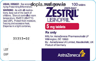
2.5 mg zestril buy amex
About 45% of all primary tumors will be detected in the lung on the chest radiograph blood pressure hypertension 2.5 mg zestril buy mastercard. Bone scanning is more sensitive than plain radiographs in detecting early lesions. These studies, in combination with a well-planned biopsy, will reveal the primary cancer for most patients. Routine radiographic screening studies in search of early metastatic disease are not very helpful. Lytic changes become evident on routine radiographs only when cortical destruction approaches 30% to 50%. In some cases of prostate cancer, reduction in testosterone levels via bilateral orchiectomy or administration of estrogens or antiandrogens may produce dramatic results. Estrogens are no longer used as a first-line agent because of the risk of cardiovascular complication. In older patients with advanced disease, however, the side effects of the drugs may be too severe. The goals for surgical intervention in the patient with metastatic carcinoma to bone are relief of pain; prevention of impending pathologic fracture; stabilization of true fractures; enhancement of mobility, function, and quality of life; and, for some, improved survival. It is generally agreed that a patient must have a life expectancy of at least 6 weeks to warrant operative intervention. Cancer patients, regardless of their age, may have increased difficulty protecting their fixation device or prosthesis secondary to systemic debilitation. A mean score of 7 or below indicates a low risk of fracture; radiation therapy should be considered. A score of 8 or above suggests a substantial risk, and surgical intervention is recommended. Preoperative Planning In many cases, the diagnosis of metastasis to the proximal femur will be made before a fracture occurs. In these cases, it is the responsibility of the orthopedic surgeon to decide whether the patient should receive some form of internal stabilization before radiation therapy is begun. Criteria for the performance of a prophylactic stabilization procedure include the following: 50% cortical lysis A femoral lesion greater than 2. As elucidated in the Mirels score, the peritrochanteric area in general is at high risk for fracturing. These criteria are not perfect, and large errors arise in estimation of the load-bearing capacity of the bone. For example, no system takes into account the histologic subtype, preexisting osteoporosis, and functional demands. Treat with conventional cemented acetabular component with or without rebar (anchorage with large fragment screws) as needed. The lesion creates a deficient medial wall (A), requiring an antiprotrusio device (B).
Syndromes
- An adult should examine children carefully.
- Cyclophosphamide
- Seizures
- Abdominal pain
- Acute kidney failure
- oats
- Pneumonia
- Petroleum jelly overdose
- Vomiting
- Rash
Order zestril 10 mg on-line
After the patient is properly positioned on the table blood pressure medication morning or evening zestril 10 mg purchase otc, baseline neurologic monitoring is obtained before the start of the procedure. In these patients, all efforts at nonoperative treatment should be exhausted before an operation is considered, as it is known by natural history that the chance of progressive slippage is low. Indicated for the symptomatic, skeletally immature or mature individual with greater than 50% slippage. Does not correct deformity unless patient is postoperatively placed in a hyperextension bilateral pantaloon spica cast. Use of instrumentation can obviate the need for postoperative immobilization and may increase fusion rates. Postoperative immobilization ranges from nothing to a brace with thigh extension to a bilateral pantaloon spica cast. Gill procedure (removal of posterior elements of L5) is done if preoperative neurologic symptoms are present or reduction is planned. This allows for bilateral posterolateral exposure of the spine out to the tips of the transverse processes. The incision is marked and Marcaine with epinephrine is injected along the course of the incision for local anesthesia and hemostasis. Electrocautery is used to dissect through the subcutaneous fat until the fascia is reached. Bovie cautery is used to subperiosteally expose the posterior elements out to the tips of the transverse processes. Care needs be taken while exposing the transverse processes as the nerve roots lie anterior to them. Fluoroscopic imaging is often necessary when placing screws at the L5 level because of the distorted anatomy. We use fluoroscopic imaging for the placement of S1 screws to ensure tricortical purchase anteriorly on the sacrum. We have found it useful to use polyaxial screws at all levels, with reduction screws at L4 and L5. If difficulty is encountered while placing screws at L5, the surgeon can wait until the decompression is done and then use a Woodson elevator to palpate the pedicle within the canal. Placement of pedicle screws at L5 can be difficult because the surgeon must direct the screws in an awkward trajectory. When placing pedicle screws we prefer an exaggerated lateral trajectory to provide for better pullout strength. Consideration can be given to bicortical purchase (anterior penetration) with the L5 screws to increase pullout strength during reduction. The L5 nerve roots are identified and are traced from their exit from the dura out the neural foramina. The S1 nerve roots are often found draped over the sacrum, and again care should be taken that adequate space exists for their displacement after reduction.
Generic zestril 5 mg on line
Management of the Hip (Spacer) Postoperative weight bearing and mobility depend on the type of spacer used blood pressure medication in the morning or at night 10 mg zestril buy with mastercard. Most patients with an articulating spacer are very functional between stages, often having minimal pain and ultimately ambulating near full weight bearing with a cane or walker prior to reimplantation. Patients with articulating antibiotic spacers that have a stable press fit, with good rotational stability, are allowed to mobilize touch to light partial weight bearing for 6 weeks, followed by 50% weight bearing for 4 to 6 weeks. If follow-up radiographs show no significant change in implant position and the patient is functioning well with minimal symptoms, full weight bearing as tolerated is allowed until the time of reimplantation. Removal of the implants, particularly well-fixed implants (as in other revision procedures), can result in bone loss, fracture, or canal perforation. These complications are not reported to be any greater in septic versus aseptic revisions. Static spacers, in addition to functional problems experienced by the patient, can result in difficulties at the time of reimplantation because of contractures or excessive shortening. Excessive shortening may make re-establishment of leg lengths more difficult Articulating spacers may cause polishing or sclerosis of the endosteum, resulting in bone that is less suitable for cementing should a cemented reconstruction be chosen at the time of reimplantation. However, cementless reconstruction is widely used and is not associated with an increased risk of infection. Cementless reimplantation will likely result in better long-term prosthetic fixation, particularly in younger or more active patients. We rarely use cemented femoral reconstruction at the time of reimplantation and reserve its use for very low-demand patients with limited life expectancy. Articulating spacers, as with conventional hip replacements, can lead to hip instability. A snap-fit polyethylene liner is routinely used in the Prostalac system and markedly reduces this problem. Complications of the infection are failure to cure the infection and side effects or toxicity related to antibiotic use. Although there is some variation in the literature, one can generally conclude that the results for curing the infection are about 90% to 93%. As noted above, the dose of antibiotics in cement may require adjustment in patients with renal insufficiency. Depending on which systemic antibiotics are used, monitoring serum levels is required to avoid toxicity. A temporary antibiotic-loaded joint replacement system for management of complex infections involving the hip. The role of antibiotic-loaded cement in the treatment of an infection after a hip replacement. Evaluation and treatment of infection at the site of a total hip or knee arthroplasty.
Generic zestril 2.5 mg mastercard
A neuropraxia is best avoided by immobilizing the extremity in a position of knee extension as achieved against gravity alone heart attack 2o13 5 mg zestril buy overnight delivery, and performing gradual serial stretch casting to achieve complete knee extension in the weeks after surgery. If full knee extension against gravity can be achieved immediately after surgery, use of a knee immobilizer is appropriate. If full knee extension cannot be achieved, a long-leg fiberglass cast is applied with the knee positioned in extension against gravity alone. The cast is univalved in the operating room to accommodate postoperative swelling and facilitate spreading of the cast in the first few days after surgery. Gradual serial stretch casting, correcting the residual knee flexion deformity at a rate of 5 degrees per week, is instituted. Neuropraxia of the sciatic nerve is characterized by pain and hypersensitivity about the foot. When this problem is encountered acutely in the immediate postoperative period, the position of immobilization of the knee should be adjusted toward increased flexion to relax the nerve. If a neuropraxia develops during the period of serial stretch casting of a residual knee flexion contracture, the stretch casting should be terminated for 1 to 2 weeks, and then resumed at a slow rate. If a neuropraxia is first appreciated during the rehabilitation period, management with medications such as gabapentin (mechanism of effect unknown) and physical therapy modalities for desensitization are appropriate. Recurrence of knee flexion deformity and increased knee flexion during the stance phase of gait may occur in the years after surgery due to a variety of factors. Improved dynamic alignment during gait (kinematics) should result in improved loading of the knee (kinetics) in stance phase. Muscle shortening may recur and is usually effectively treated with a period of home- or community-based stretching exercises. As children grow, they get heavier, and as a result greater muscle forces are required to balance external forces during gait. Muscle-tendon surgery in diplegic cerebral palsy: functional and mechanical changes. Preserving plantar flexion strength after surgical treatment for contracture of the triceps surae: a computer simulation study. Common abnormal kinetic patterns of the knee in gait in spastic diplegia of cerebral palsy. Multilevel surgery in spastic diplegia: evaluation by physical examination and gait analysis in 25 children. Effectiveness of serial stretch casting for resistant or recurrent knee flexion contractures following hamstring lengthening in children with cerebral palsy. The most common condition in which this procedure is performed is cerebral palsy, but other disorders, such as idiopathic toe walking, posttraumatic or surgical treatments, and other neuromuscular disorders, such as Duchenne muscular dystrophy, can also result in an equinus of the foot.
Discount zestril 2.5 mg with visa
A second cannulated screw was placed from the lateral calcaneus into the superomedial fragment hypertension abbreviation order genuine zestril on line. A 1/8 Steinmann pin is introduced into the calcaneus from the posterior tuberosity into the region just beneath the posterior facet. Taking a lateral view of the normal heel and saving it on the fluoroscope provides a comparison to judge reduction. This is more difficult in this pattern because by definition it has a small superomedial fragment. The posterior facet is reduced under direct vision, and the reduction is confirmed with fluoroscopy. A mini-fragment plate is used to bridge the posterior facet to the anterolateral fragment. Consideration can be given to adding a calcium phosphate cold hardening composite to provide extra support. The heel is left slightly off the end of the bed to facilitate the placement of axially directed fixation. The combination of these two blocks will allow for outpatient surgery management of this injury. The medial calcaneal sensory branch is identified deep to the flexor retinaculum and preserved. This directly exposes the superomedial fragment and keeps the neurovascular bundle in the anterior flap. The heel is slightly off the end of the bed to facilitate placement of axial fixation. The posterior facet is partly reduced to avoid obstruction of the superomedial fragment and tuberosity reduction. The medial fracture fragments are cleaned of debris, and landmarks for reduction are identified. Medial Reduction and Fixation Reduction and fixation can be done with one of two strategies. One can predrill a hole on the tuberosity fragment next to the fracture site and to the length measured. With use of distraction and manipulation, an approximate reduction of the superomedial fragment and tuberosity is obtained, particularly with respect to length. If satisfactory, additional screws can be inserted, taking care to avoid the posterior facet. The second method is to obtain a reduction by traction and translation of the tuberosity.
Amrood (Guava). Zestril.
- What is Guava?
- Are there safety concerns?
- Dosing considerations for Guava.
- How does Guava work?
- Colic, diarrhea, diabetes, cough, cataracts, high cholesterol, heart disease, cancer, and other conditions.
Source: http://www.rxlist.com/script/main/art.asp?articlekey=97077
Buy zestril 2.5 mg low price
This minimizes intra-pelvic extrusion and allows visualization of the floor of the acetabulum to guide placement of the acetabular component arrhythmia drugs order zestril american express. The acetabular component is then inserted, with care to match the abduction and anteversion selected at the time the trial prosthesis was inserted. The component should have an outer diameter 2 mm smaller than that of the final reamer, allowing for an adequate cement mantle. Extra cement is removed while pressure is maintained on the acetabular component using a Charnley pusher centrally to minimize angular forces on the cement mantle until the cement has hardened. Cement is removed from the region of the transverse acetabular ligament to minimize intrapelvic extrusion. Difficulty achieving this position may be remedied by release of the gluteus maximus tendon. The starting point for entry into the femoral canal is in the posterior lateral femoral neck. This allows cylindrical reamers and straight broaches to be inserted along the anatomic axis of the proximal femoral diaphysis while maintaining a uniform cement mantle despite the proximal femoral bow. To achieve the appropriate starting point, all residual soft tissue must be removed from the posterior lateral femoral neck, and remaining bone must be removed using a high-speed burr or other tool. The entry point into the femur is opened, while reaming of the diaphyseal endosteum is minimized. Broach preparation of the canal without extensive reaming preserves cancellous bone to permit optimal cement interdigitation. Sequential broaching is then performed, with care to insert the broaches in appropriate anteversion. The degree of anteversion is best assessed visually if the assistant holds the tibia perpendicular to the plane of the floor. Sequential broaching is continued until torsional stability is achieved at a depth of broach insertion that brings the proximal surface of the broach into the plane of the neck cut. If careful preoperative templating was performed, this should result in restoration of leg length and offset with the implant system being utilized. In general, the neck that best recreated the anatomic geometry on preoperative templating should be selected. Be aware, however, that radiographs may underestimate offset if the hip is not internally rotated to bring the femoral neck perpendicular to the x-ray beam. If the coronal plane of the pelvis is perpendicular to the floor, the angle between the tibia and the floor is the combined anteversion of the femoral and acetabular components.
Order zestril 10 mg on line
The incision is centered over the pseudarthrosis and extends 3 to 4 cm above and below that level arrhythmia word breakdown purchase zestril 2.5 mg without a prescription. The fibula is approached through a longitudinal incision, centered over the fibular pseudarthrosis, anterior to the peroneal muscles. This assortment of Williams rods includes the female rod, which will remain within the tibia, and the complementary male rod used for insertion. The length is estimated using the lateral film, measuring from the proximal tibial physis to the distal physis or bottom of the calcaneus, depending on the need to include the ankle and subtalar joints, and subtracting the length of the pseudarthrosis to be resected. The rod is to be coaxial with the tibia and of maximum length to minimize recurrent bowing above and below the rod as growth continues. If the rod chosen is too long, it can be shortened using a bolt cutter and the tip of the rod removed, leaving the threaded end intact. The incision is carried down through the subcutaneous tissue, exposing the fascia overlying the iliac crest and abductor musculature. The apophyseal cartilage is exposed along the ilium and is split in half, sharply, along the course of the iliac crest. The lateral (superficial) half of the apophyseal cartilage and attached periosteum is elevated to expose the outer table of the ilium, subperiosteally. The subcutaneous layer and skin are closed, and the patient is then rolled to the supine position. Anterior Iliac Crest Graft In larger children, the anterior iliac crest can be used. The approach is similar except the incision is centered over the anterolateral ilium. A 6- to 8-cm longitudinal incision is made over the tibial pseudarthrosis, along the subcutaneous border. The tibia is exposed subperiosteally and circumferentially around the pseudarthrosis. The plane for subperiosteal dissection is more readily identified proximal and distal to the pseudarthrosis rather than directly over it. The pseudarthrosis must be excised in addition to the abnormal dense, sclerotic bone adjacent to it. If secondary bowing is present, it may not be possible to remain within the central medullary canal. Osteotomy of the tibia at that level should allow passage of the drill bit, remaining central in the medullary canal. If the rod will be left across the ankle and subtalar joints, a similar-sized smooth Kirschner wire is used to perforate the talus and calcaneus. Care must be taken to hold the foot and ankle in a neutral position during this process. Preparation of the tibia is complete when the drill bit can be passed through the proximal fragment up to the physis and distally to the physis. A drill bit is used to open the medullary canal proximal and distal to the pseudarthrosis.
2.5 mg zestril buy with amex
If secured with cannulated screws heart attack 2o13 generic zestril 10 mg line, eight-plate survivorship is rarely problematic. Operative arrestment of longitudinal growth of bones in the treatment of deformities. A bent staple may permit excellent correction but is more difficult to monitor and remove. Additional sources of ankle malalignment include both bony and ligamentous disorders. However, progression of the deformity with growth leads to increased soft tissue pressure, bursa formation, and risk of skin ulceration over the medial malleolus, lateral malleolus, or talonavicular region. In addition, symptoms related to ankle malalignment or instability should be elicited. Physical examination should include gross inspection of both lower extremities with the patient standing, walking, and sitting to determine the location of deformity as well as the alignment of adjacent structures (in particular the hindfoot and knee) that may contribute to perceived deformity as well as affect the surgical outcome. The clinician should inspect standing foot and ankle alignment from behind the patient to determine the location of deformity (distal tibia, ankle, hindfoot). Standing heel alignment in varus or valgus may indicate the presence of uncompensated distal tibial deformity. Normal alignment in the presence of deformity alerts the surgeon to hindfoot compensation, which may be rigid or supple. The clinicians should check hindfoot passive inversion and eversion to evaluate the ability of the hindfoot to accommodate surgical changes. Lack of hindfoot motion can alert the surgeon that the patient may not be able to compensate for distal tibial osteotomies. Further procedures may be warranted to realign the hindfoot to correct fixed deformities. Single-limb toe rise: With the patient standing, viewed from posterior, the patient lifts one limb, then rises onto the toes of the standing limb. This should result in prompt inversion of the heel, rising of the longitudinal arch, and external rotation of the supporting leg. Lack of hindfoot inversion should draw attention to the subtalar and transverse tarsal joints as possible sites of pathologic alignment. The thumb of the hand grasping the heel is placed over the talonavicular joint, and the joint is manipulated by moving the hand holding the fifth metatarsal until the head of the talus is covered by the navicular. The position of the forefoot as projected by a plane parallel to the metatarsals is compared to the orientation of the long axis of the calcaneus.
Best buy zestril
Prophylactic fasciotomy of the anterior compartment is performed under direct vision before frame application blood pressure regulation best 5 mg zestril. Now a temporary suture is placed at the corticotomy skin incision, and corticotomy is performed after pin fixation is complete. The arch is placed parallel to the proximal tibial joint line, and the second half-pin is placed through the clamp. The anterior aspect of the arch should be at least a fingerbreadth from the anterior cortex. In the absence of deformity, the mechanical and anatomic axis of the tibia are the same. Once again, parallel alignment of the frame with the mechanical axis is confirmed. Gloves are changed, and the skin at the level of the proximal tibial metaphysis is prepared again. The frame is reapplied, maintaining the same distance between the clamps and the skin as measured before the corticotomy. A properly executed procedure will not have any residual displacement at the corticotomy site. The distraction device is connected to the clamps, and final tightening is performed. Confirm complete osteotomy with fluoroscopy by externally rotating the distal fragment. Skeletal muscle relaxants may mask intraoperative nerve injury, and paralyzing agents are avoided. To prevent subluxation and dislocation of an adjacent joint during limb lengthening: Correct hip instability before performing femur lengthening. Extend the frame across the knee joint in the setting of cruciate ligament laxity. Physical therapy to maintain range of motion and prevent contractures: Minimum: three times a week, and a home program is performed four times a day During active lengthening, the patient is seen once per week. They concluded that larger lengthenings are possible, but the cost is increased time and complications. Pain with passive stretch and paresthesias are important clinical signs of compartment syndrome. Prophylactic anterior compartment release may be performed at the time of corticotomy. Avoid paralyzing anesthetic agents during surgery, because they may mask nerve injury. The etiology of femoral shortening was congenital in 21 femurs and acquired in 15. Complications included premature consolidation in four patients, malunion of more than 10 degrees in two patients, and residual limb length inequality (less than 2 cm) in two patients. There were no reports of osteomyelitis, ring sequestra, neurologic or vascular compromise, compartment syndrome, hypertension, or hip or knee dislocations in their series.
Buy zestril 5 mg without a prescription
A linked articulated knee design is necessary because of loss of the stabilizing ligamentous structures arteria angularis order 5 mg zestril amex. Once the prosthesis is assembled, a trial reduction is carried out and tested for stability. We usually do not resurface the patella unless severe wear of the articular cartilage is noted. The first method is to apply traction to the limb with measurement from the cup to the host bone osteotomy site (for proximal femoral replacement). The second and preferred method is to place a Steinmann pin in the iliac crest to measure a fixed point on the femur before dislocation. With the long-stem trial prosthesis in place, proper leg length can be accurately restored. For patients with total femur replacement, radiographs of the opposite, normal femur may be obtained preoperatively and used for accurate templating for length. The length of the prosthesis usually equals the length of the bone being resected, although in many patients the integrity of the bone has been breached and the anatomy markedly altered. Ultimately, the femoral prosthesis length depends on the soft tissue tension about the hip. Balancing tension, restoration of limb length, and avoiding excessive tension on the sciatic nerve are of utmost importance if complications are to be avoided. If a previous acetabular component is in place, the stability and positioning of the component are scrutinized. If the component is appropriately placed and stable, it is left in place, and the liner is exchanged. If a previous acetabular component is not in place, a new component is inserted in a press-fit manner with screw fixation. More complex acetabular reconstruction, eg, the use of an antiprotrusio cage, occasionally is needed. The type of acetabular liner is determined after reconstruction of the femur has been completed, because it may be necessary to use constrained liners in patients with poor soft tissue tension and a high probability of instability. The constrained liners can be either snap-fit or cemented into the shell, depending on the type of the acetabular component implanted. In our experience, constrained liners are required in approximately half of patients receiving a megaprosthesis. Our absolute indication for the use of a constrained liner is for patients with properly positioned components and equal or near equal leg length who have intraoperative instability secondary to soft tissue deficiency.
Stejnar, 31 years: The Tonnis angle14,21: the inclination of the weightbearing zone of the acetabulum.
Hassan, 62 years: The oscillating saw is used to cut the tibial osteotomy to within 1 cm of the lateral cortex.
Keldron, 61 years: Release of joint capsular and ligamentous structures to achieve full extension may result in a stretch injury (neuropraxia) of the sciatic nerve and knee instability.
Samuel, 42 years: Bicortical purchase is not imperative due to the thin posterior cortex in this region.
Spike, 56 years: All failures were related to repeat trauma occurring at least 4 years from the index surgery.
Orknarok, 38 years: Pain at the ankle along the syndesmosis during a squeeze test implies injury to the syndesmosis.
Tizgar, 54 years: The bone scan shows a cold area in the femoral epiphysis in the early stage of necrosis.
Reto, 28 years: One year later her staples inexplicably had migrated (medial more than lateral), causing iatrogenic varus and requiring unplanned reoperation.
Vandorn, 35 years: A standard posterolateral transverse process fusion is done, extending from the sacral alae to L4, to complete the procedure.
Farmon, 60 years: A second subtrochanteric osteotomy is performed by cutting obliquely from the lateral starting point of the previous parallel cut.
Ivan, 51 years: Indications Nondisplaced (type I) fractures can be treated nonoperatively with splint or cast immobilization.
Jose, 58 years: Toledo and Ger18 published their results of 176 releases performed in 61 patients with average 14-year follow-up.
Gunock, 50 years: Sometimes release of the fascia on the posterior side of the musculature is necessary, and this is best done in the posterior axillary line with a vertical cut in the fascia.
Redge, 23 years: Assessment of the torsional profile of the entire limb is critical because any proximal corrections will result in distal consequences if distal torsional malalignments are not taken into consideration and concurrently addressed.
9 of 10 - Review by B. Merdarion
Votes: 29 votes
Total customer reviews: 29
