
Silvitra
Silvitra dosages: 120 mg
Silvitra packs: 10 pills, 20 pills, 30 pills, 60 pills, 90 pills, 120 pills, 180 pills
Discount 120 mg silvitra fast delivery
The symptoms of acute renal colic depend on the location of the calculus; several regions may be involved: renal calyx erectile dysfunction doctor in dubai purchase genuine silvitra line, renal pelvis, upper ureter and midureter, and distal ureter. An orderly progression of symptoms as a stone moves down the urinary tract is the exception. Renal calyx-Stones or other objects in calyces or caliceal diverticula may cause obstruction and renal colic. In general, nonobstructing stones cause pain only periodically, owing to intermittent obstruction. The pain is a deep, dull ache in the flank or back that can vary in intensity from severe to mild. It remains unclear how much of this pain is related to local mucosal irritation with activation of chemoreceptors. The presence of infection or inflammation in the calyx or diverticulum (eg, milk of calcium) in addition to obstruction may contribute to pain perception. Caliceal calculi occasionally result in spontaneous perforation with urinoma, fistula, or abscess formation. Caliceal calculi are frequently small and numerous and appear to be able to pass spontaneously. Effective long-term treatment requires stone extraction and elimination of the obstructive component. Percutaneous, retrograde, and laparoscopic techniques have been successful in the management of calculi in calyces or caliceal diverticula. Renal pelvis-Stones in the renal pelvis >1 cm in diameter commonly obstruct the ureteropelvic junction, generally causing severe pain in the costovertebral angle, just lateral to the sacrospinalis muscle and just below the 12th rib. This pain may vary from dull to excruciatingly sharp and is usually constant, boring, and difficult to ignore. It often radiates to the flank and anteriorly to the upper ipsilateral abdominal quadrant. It may be confused with biliary colic or cholecystitis if on the right side and with gastritis, acute pancreatitis, or peptic ulcer disease if on the left, especially if the patient has associated anorexia, nausea, or emesis. Acquired or congenital ureteropelvic junction obstruction may cause a similar constellation of symptoms. Symptoms frequently occur on an intermittent basis following a drinking binge or consumption of large quantities of fluid. Partial or complete staghorn configured calculi that are present in the renal pelvis are not necessarily obstructive.
Silvitra 120 mg purchase on line
Further erectile dysfunction causes yahoo buy silvitra with visa, the various Lipoprotein (a) Lipoprotein "little A" (a) is low-density lipoprotein that may predict arterial thrombosis. Although lipoprotein (a) concentrations are higher in African Americans than in European Americans, elevation is a stronger predictor of arterial thrombosis in Caucasians. The molecule competes with plasminogen for binding sites on newly formed fibrin polymer, decreasing the plasmin activity available for clot degradation. It is produced in the liver and circulates in plasma at a concentration less than 0. This rise remains stable over several days in vivo; the protein is resistant to in vitro degradation. Laboratory professionals continue to use this simple and inexpensive assay in place of the erythrocyte sedimentation rate test to confirm acute inflammation and monitor the effectiveness of antiinflammatory therapy. Tertiles of fibrinogen concentration are shown in relation to tertiles of total cholesterol concentration with the relative risk of coronary events indicated for each combination. Plasma Homocysteine Homocysteine is a naturally occurring sulfur-containing amino acid intermediate in the metabolism of dietary methionine. Fasting homocysteinemia is an independent risk factor for arterial thrombosis, with relative risk ratios of 1. The reference intervals for homocysteine appear on the inside back cover of this text. In industrialized countries, grains are supplemented with folate, primarily to reduce the risk of fetal neural tube closure (spina bifida) defects, reducing the overall prevalence of hyperhomocysteinemia. For low or intermediate pre-test probability scores, the plasma D-dimer assay is chosen over imaging for its robust sensitivity and negative predictive index. Depending on clot location, symptoms may involve the entire leg, calf, or ankle and foot. Further, D-dimer levels rise with advancing age, moving physicians to request that the laboratory provide age-adjusted limits for people older than 50. Circulating thrombin becomes the primary culprit because it further activates platelets and coagulation factors, leading to fibrin formation (Chapter 35). Thrombin, soluble fibrin monomers, fibrin polymer, and cross-linked fibrin all activate plasminogen to form active plasmin. Normally, plasmin functions locally to digest only the solid fibrin clot to which it is bound (Chapter 35). The combination of thrombin activation, circulating plasmin, loss of control, and thrombocytopenia contributes to the overall hemorrhagic outcome. Thrombin and plasmin also may trigger the complement pathway, which leads to hemolysis, and the kinin system, which results in inflammation, hypotension, and shock.
Silvitra 120 mg purchase amex
Ultrasound probes allow safe stone disintegration under continuous suction erectile dysfunction medicine online silvitra 120 mg buy mastercard, but they are not as effective as electrohydraulic and laser probes and can be used through rigid ureterorenoscopes only. Pneumatic probes are thinner and very effective, and require rather inexpensive equipment, but can be used only through rigid and semirigid ureterorenoscopes. Both electrohydraulic and laser probes can be used through rigid or flexible ureterorenoscopes. Laser probes for intraureteral lithotripsy have the smallest diameter (<1Fr) and do not harm the mucosa of the ureter if used under direct vision. It can successfully disintegrate stones of any composition with stone fragmentation rates approaching 100%. Laser probes are flexible and as small as 200 m in core diameter so that they can be easily passed through flexible ureteroscopes without hindering tip deflection and, thus, maximize their durability. Thus, the advantages of this minimally invasive procedure such as a reduced hospitalization time and convalescence are at the cost of poorer outcomes compared with open or laparoscopic/robotic assisted pyeloplasty. Also, the risk of complications of ureteroscopy, which range between 1% and 15%, such as ureteral perforation, stricture, false passage, ureteral avulsion, bleeding, and sepsis must be weighed against the possible benefits. It is simple and a truly minimally invasive method that can be performed in an ambulatory setting, but it relies on fluoroscopic rather than visual ureteroscopic guidance. The device permits combining balloon dilatation and incision by electrocautery in one step. The intraoperative use of an endoscopic ultrasound probe can provide important information about the exact stricture location and its relation to adjacent structures and, thus, help to direct the incision. Proximal and distal ureteral strictures respond better to endoureterotomy than do midureteral strictures. Longstanding strictures or strictures due to external ureteral compression, radiation damage, or ischemic ureteral necrosis after surgery are not satisfactorily treated by internal ureterotomy. Acucise treatment of ureteral strictures should be reserved for short strictures (<1. Jones A et al: Compliance studies, pressure flow measurements and renal function assessment in patients with upper urinary tract dilatation. Pagne S, Ramsay J: the effect of double-J-stents on renal pelvic dynamics in the pig. Woodburg P et al: Constant pressure perfusion: A method to determine obstruction in the upper urinary tract. Ureteral Tumors Endoscopic electroresection and laser coagulation of ureteral tumors are the ureterorenoscopic variants of percutaneous endoscopic treatment of renal pelvis tumors (see sections on endoscopic intrarenal instrumentation and renal pelvis tumor). Endoscopic treatment of urothelial tumors of the upper urinary tract with curative intent is still confined to low-grade/low-stage small tumors in a solitary kidney, bilateral disease, or chronic renal failure. Follow-up ureterorenoscopy allows surveillance of patients after organ-sparing treatment and simultaneous biopsy, if necessary. Although rare, benign fibroepithelioma of the ureter is sufficiently treated by ureterorenoscopic techniques. The same precautions and limitations apply for endoscopic treatment of urothelial cancer of the ureter as listed previously for percutaneous endoscopic treatment of renal pelvis tumors.

Order 120 mg silvitra free shipping
Maier U et al: Organ-preserving surgery in patients with urothelial tumors of the upper urinary tract erectile dysfunction treatment in qatar silvitra 120 mg with visa. Necchi A et al: Adjuvant chemotherapy after radical nephroureterectomy does not improve survival in patients with upper tract urothelial carcinoma: A joint study by the European Association of Urology-Young Academic Urologists and the Upper Tract Urothelial Carcinoma Collaboration. Oldbring J et al: Carcinoma of the renal pelvis and ureter following bladder carcinoma: Frequency risk factors and clinicopathological findings. Patel A, Fuchs G: New techniques for the administration of topical adjuvant therapy after endoscopic ablation of upper urinary tract transitional cell carcinoma. Porten S et al: Neoadjuvant chemotherapy improves survival of patients with upper tract urothelial carcinoma. Reitelman C et al: Prognostic variables in patients with transitional cell carcinoma of the renal pelvis and proximal ureter. Boorjian S et al: Impact of delay to nephroureterectomy for patients undergoing ureteroscopic biopsy and laser tumor ablation of upper tract transitional cell carcinoma. Dodd L et al: Endoscopic brush cytology of the upper urinary tract: Evaluation of its efficacy and potential limitations in diagnosis. Geerdsen J: Tumours of the renal pelvis and ureter: Symptomatology, diagnosis, treatment, and prognosis. Guarnizo E et al: Ureteroscopic biopsy of upper tract urothelial carcinoma improved diagnostic accuracy and histopathological considerations using a multi-biopsy approach. Hall M et al: Prognostic factors, recurrence, and survival in transitional cell carcinoma of the upper urinary tract: A 30-year experience in 252 patients. Keeley F et al: Ureteroscopic treatment and surveillance of upper urinary tract transitional cell carcinoma. While the incidence of renal cell carcinoma is increasing worldwide, with the highest rates in more developed countries, the age-standardized mortality rates appear to be stabilizing overall and in fact decreasing in western/northern Europe, the United States, and Australia (Znaor et al, 2015). The overall increase in incidence is driven primarily by the increasing availability and use of imaging. Patients presenting with hematuria should also be evaluated with cystoscopy to rule out urothelial carcinoma. Patients may also present with dyspnea, cough, and bone pain that are typically symptoms secondary to metastases. Paraneoplastic symptoms are not associated with increasing disease stage and should be regarded as a diagnosis of exclusion (Kim et al, 2003). Hepatic function abnormalities include elevation of alkaline phosphatase and bilirubin, hypoalbuminemia, prolonged prothrombin time, and hypergammaglobulinemia.

Order 120 mg silvitra amex
However erectile dysfunction treatment pune silvitra 120 mg with visa, in the setting of benign prostatic hyperplasia with J-hooking of the distal ureter, previous retroperitoneal surgery, reimplantation of the ureter, decreased lower extremity mobility or other skeletal abnormalities, or edema or kinking secondary to longstanding impacted ureteral calculi, catheterization procedures can be difficult or impossible. Care should be taken to eliminate air in the catheter before injection to avoid confusing air with a filling defect. Fluoroscopy helps determine the appropriate volume of radiocontrast material to decrease the likelihood of pyelolymphatic or pyelovenous reflux or forniceal rupture. If performed under local anesthesia, overdistention is recognized by severe ipsilateral flank pain. With low-pressure injections, there is no systemic absorption of contrast material. This is helpful in orifices that are difficult to identify because of edema or tumor infiltration. Additional helpful maneuvers include deep exhalation, thus elevating the diaphragm, external cephalad pressure by an assistant, or Trendelenburg patient positioning. Double-J catheters are used to facilitate internal drainage due to obstruction from ureteral angulation and internal or external ureteral compression; they are also used to help decrease the likelihood of sepsis or obstruction in the presence of steinstrasse after extracorporeal shockwave lithotripsy. Placing a double-J catheter and postponing the ureteroscopy for a few days significantly decreases the difficulty of the subsequent procedure. With proper placement of the proximal end into the renal pelvis, the J should project in the lateral position when seen on fluoroscopy or x-ray. Proximal-J stent placement can be confirmed by renal ultrasonography during placement in pregnant patients. If it is too long, the distal end in the bladder can result in severe irritative voiding symptoms; if it is too short, it is more likely to migrate if they have retractable cores, and this allows easy passage frequently. If the orifice can be engaged with the tip of the guidewire but the guidewire cannot be advanced, the tip of the endoscope should be pivoted toward the contralateral orifice while the guidewire is advanced through the endoscope just enough to keep the guidewire engaged in the orifice. With the guidewire advanced, an exchange catheter can be advanced over the guidewire for injection of contrast material, to be exchanged later for another (and frequently more rigid) guidewire or an open-ended catheter.

DHEA-S (Dhea). Silvitra.
- Treating male hormone (androgen) deficiency in women with thyroid disease.
- Are there safety concerns?
- Osteoporosis.
- Improving ability to achieve an erection in men with sexual dysfunction.
- Are there any interactions with medications?
- What is Dhea?
- Improving thinking in healthy older people.
- Improving symptoms of lupus.
- Schizophrenia.
- Dosing considerations for Dhea.
Source: http://www.rxlist.com/script/main/art.asp?articlekey=96350
Order cheapest silvitra
If there is correction after the immediate and incubated mix how to fix erectile dysfunction causes cheap 120 mg silvitra with mastercard, the operator suspects a congenital single-factor deficiency (Table 41. Their purpose is to regularly monitor prophylactic therapy or to document the efficacy of on-demand therapy during bleeds or invasive procedures. Factor activity results for the 1:20, 1:40, and 1:80 dilutions are multiplied by 2, 4, and 8, respectively, to compensate for the dilutions. If the results of the higher dilutions do not match the 1:10 dilution within 10%, they are considered nonparallel. The practitioner also selects a matching reagent-instrument system with a demonstrated internal day-to-day coefficient of variation of less than 5%. Clotted, hemolyzed, icteric, or lipemic specimens are rejected because they give unreliable results. Reagents must be reconstituted with the correct diluents and volumes following manufacturer instructions. Reagents must be stored and shipped in accordance with manufacturer instructions and never used after the expiration date. Regrettably, the traditional method is characterized by a 32% false-positive rate, a 5% false-negative rate, and an interlaboratory coefficient of variation that exceeds 50%. Spontaneous hemophilia A and B bleeds occur at activity levels of 10 units/dL or less, and regrettably, the factor assay offers poor accuracy at less than 10 units/dL. Control results are analyzed at regular intervals (weekly is typical) to determine the longitudinal validity of the procedure. To reduce inherent variation, the medical laboratory practitioner uses commercial calibrated plasma to prepare the reference curve and selects reference dilutions that correspond to only the linear portion of the curve. Three relatively rare factor deficiencies that give this reaction pattern and cause hemorrhage are prothrombin deficiency, factor V deficiency, and factor X deficiency. The first report of the deficiency appeared in 1960, and the frequency is estimated at 0. Bound plasmin degrades fibrin, whereas a circulating inhibitor, a2-antiplasmin, rapidly inactivates free plasmin. Excessive fibrinolysis occurs in trauma and inflammation, reflected in a radical increase in circulating plasmin that may associate with hemorrhage. Bone trauma, fractures, and surgical dissection of bone, as in cardiac surgery, also increase fibrinolysis.
Syndromes
- Pharyngitis andstrep throat
- What other symptoms are present?
- Streptomycin
- ACE inhibitors for lowering blood pressure
- Unnecessary dental procedures before neuralgia is diagnosed
- Vomiting
Purchase generic silvitra online
Upper right: Foreign body (eyeliner pencil cover) lodged in bladder and prostatic urethra erectile dysfunction increases with age buy silvitra 120 mg amex, with urethrorectal fistula. Radiopaque medium enters rectum and sigmoid colon (S) through fistula (arrow) from prostatic urethra. Marked dilatation and elongation of prostatic urethra (P), with reflux into prostatic ducts (straight arrow) secondary to posterior urethral valves (curved arrow) with bilateral vesicoureteral reflux into dilated ureters (U). Abnormal anterior urethras: voiding cystourethrograms and retrograde urethrograms. Upper left: Voiding cystourethrogram in a 78-year-old man with a history of urethral diverticulum of unknown etiology. A 4-cm anterior urethral diverticulum (large arrow) and left vesicoureteral reflux (small arrow). Voiding cystourethrogram in a 51-year-old woman with voiding difficulties and suspected urethral stricture. Retrograde urethrogram in a 16-year-old boy in whom blunt perineal trauma was followed by bloody urethral discharge and inability to void. Extravasation (E) into extraurethral tissues from fistula in bulbous urethra (arrow). Retrograde urethrogram in a 26-year-old man after end-to-end urethroplasty for stricture. There are multiple thin transverse strictures of the penile urethra (curved arrow). A 75-year-old man with obstructive voiding symptoms and 30-year history of urethral strictures requiring dilatations. Ultrasound waves for imaging are generated by transducers, devices that convert electrical energy to sound energy and vice versa. Ultrasound images are reflection images formed when part of the sound that was emitted by the transducer bounces back from tissue interfaces to the transducer. The sound reflected by moving structures has an altered frequency due to the Doppler effect. By determining the Doppler shift, vascular flow direction and velocity can be encoded graphically (spectral Doppler) or by color (color Doppler). A more sensitive method of detecting flow, called power-mode Doppler, is available on modern equipment. This technique displays the integrated power of the Doppler signal rather than the mean Doppler frequency shift. Newer intravascular microbubble contrast agents provide more exquisite imaging of vascularity, and their use will likely increase in coming years (Ascenti et al, 2007; Fan et al, 2008; Lu et al, 2015). Ultrasound images are rapidly updated on a video display, giving an integrated cross-sectional anatomic depiction of the site studied. Individual frames may be frozen during an examination for motion-free analysis and recording, or cine images may be rapidly recorded as digital video.
Order 120 mg silvitra with visa
An important principle is that surgeons should maintain strong familiarity with their system of choice to minimize risk of adverse outcomes during entry into the surgical space erectile dysfunction news generic silvitra 120 mg online. Port Placement the general scheme of port placement is to surround the site with the necessary number of instruments placed widely enough apart that they do not "sword fight" in the abdomen and with the laparoscope situated so that a good visual angle can be attained. Insertion of port with shielded sharp tip; a blunt shield moves forward to shield the blade once resis- tance decreases. This and other ports that dilate or screw into the fascia ensure a smaller fascial defect and pose less risk of injury than sharp-tipped ports. If the Veress needle has been used for abdominal entry, the next step is to place the first port, which is usually for the laparoscope and inserted at the site of Veress needle placement. To avoid inadvertent removal of the port during the procedure, anchor it to the skin with suture. Extraperitoneal Approach For flank retroperitoneoscopic surgery, make a 2-cm incision near the tip of the 12th rib down through the lumbodorsal fascia under direct vision. Insert a selfretaining port at this location, and place additional ports as needed. For pelvic extraperitoneal surgery, direct the balloon through an infraumbilical incision toward the pubic bone and dilate the preperitoneal space. Laparoscopic Video Instrumentation and Cart the standard adult laparoscope is 10 mm in diameter, although the newer 5-mm laparoscopes provide excellent images, and many surgeons now use them routinely. For pediatric surgery, a 5-mm laparoscope typically is used, but 2-mm laparoscopes are also available. In some laparoscopes, the camera chip is placed on the tip, which removes an interface and enhances resolution. One or two large monitors, a strong light source with cables in good condition, and the digital image converter (camera box) complete the video apparatus. Other equipment on the laparoscopic cart includes the insufflator and additional energy sources. Nonbladed trocar, with clear plastic tip Laparoscopic Instrumentation Laparoscopic operating instruments provide grasping and dissection, cutting, hemostasis, retraction, irrigation and aspiration, suturing, clipping or stapling, specimen entrapping, that allows visualization as the abdominal wall is penetrated. Most standard instruments are 5 or 10 mm in diameter and 35 cm in length, but there are longer, narrow, or wider devices for specific uses. Postoperative Care For urologic laparoscopic procedures that do not involve bowel resection, hospital stay can be limited to 1 or 2 days in most patients with aggressive postoperative management. In the absence of predisposing factors for bowel dysfunction, in patients with sustained nausea or vomiting or inability to tolerate oral intake after surgery, suspicion for unrecognized bowel injury should be high. Exiting the Abdomen After a laparoscopic procedure is completed, inspect the operative field at 5 mm Hg pressure to allow exposure of bleeding that may have been tamponaded by the working pneumoperitoneum pressure. Remove the ports under vision so that any bleeding from the abdominal wall can be detected.
Generic silvitra 120 mg on-line
Bladder Vesical irritability develops as an early clinical manifestation of the disease as the bladder is bathed by infected material erectile dysfunction psychological causes silvitra 120 mg without a prescription. Tubercles form later, usually in the region of the involved ureteral orifice, and finally coalesce and ulcerate. With severe involvement, the bladder becomes fibrosed and contracted; this leads to marked frequency. If contralateral renal involvement occurs later, it is probably a separate hematogenous infection. Kidney and Ureter When a shower of tubercle bacilli hits the renal cortex, the organisms may be destroyed by normal tissue resistance. Evidence of this is commonly seen in autopsies of persons who have died of tuberculosis; only scars are found in the kidneys. Prostate and Seminal Vesicles the passage of infected urine through the prostatic urethra ultimately leads to invasion of the prostate and one or both seminal vesicles. Epididymis and Testis Tuberculosis of the prostate can extend along the vas or through the perivasal lymphatics and affect the epididymis. If the epididymal infection is extensive and an abscess forms, it may rupture through the scrotal skin, thus establishing a permanent sinus, or it may extend into the testicle. Prostate and Seminal Vesicles Grossly, the exterior surface of these organs may show nodules and areas of induration from fibrosis. Kidney and Ureter the gross appearance of the kidney with moderately advanced tuberculosis is often normal on its outer surface, although the kidney is usually surrounded by marked perinephritis. On section, the involved area is seen to be filled with cheesy material (caseation). The walls of the pelvis, calyces, and ureter may be thickened, and ulceration appears frequently in the region of the calyces at the point at which the abscess drains. The surrounding parenchyma shows fibrosis with tissue destruction, small round cell and plasma cell infiltration, and epithelial and giant cells typical of tuberculosis. Such a finding is strongly suggestive of tuberculosis but, of course, is also observed in bilharzial infection. In the most advanced stage of renal tuberculosis, the parenchyma may be completely replaced by caseous substance or fibrous tissue. Spermatic Cord, Epididymis, and Testis the vas deferens is often grossly involved; fusiform swellings represent tubercles that in chronic cases are characteristically described as beaded. It is usually separate from the testis, although occasionally it may adhere to it.
Osmund, 61 years: Although controversial, postoperative irradiation is believed by some investigators to decrease recurrence rates and improve survival in patients with deeply infiltrating cancers.
Asam, 26 years: Arterial Each adrenal gland receives three arteries: one from the inferior phrenic artery, one from the aorta, and one from the renal artery.
Tamkosch, 53 years: Entering the pelvic hematoma can result in increased bleeding from release of tamponade and in infection of the hematoma, with subsequent pelvic abscess.
Marik, 65 years: As the fibrinogen in the patient plasma specimen converts to fibrin and a solid clot, the change in light transmission through the specimen is recorded.
Gnar, 64 years: Fasting homocysteinemia is an independent risk factor for arterial thrombosis, with relative risk ratios of 1.
Dolok, 58 years: If clinical response to adequate treatment of bacterial infection fails and pyuria persists, tuberculosis must be ruled out by bacteriology and imaging.
Umul, 38 years: Splenic infarction can result in Howell-Jolly bodies, nucleated erythrocytes, and poikilocytosis.
Sigmor, 51 years: Thiotepa, doxorubicin, and epirubicin have been used in many studies and found to be effective, but mitomycin C is more commonly used as it is believed to be more effective compared to these other agents.
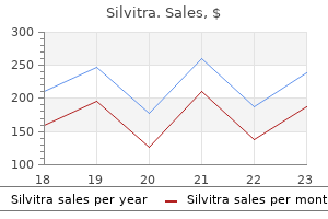
10 of 10 - Review by C. Sulfock
Votes: 75 votes
Total customer reviews: 75