Actos
Actos dosages: 45 mg, 30 mg, 15 mg
Actos packs: 30 pills, 60 pills, 90 pills, 120 pills, 180 pills, 240 pills, 360 pills, 270 pills
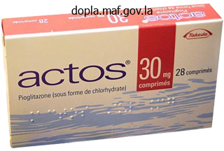
30 mg actos buy fast delivery
If the patient has an unstable pelvic fracture diabetic diet calculator 45 mg actos order with mastercard, early application of a pelvic sheet or binder is indicated and should precede additional diagnostic workup. Early application of a binder around the pelvis is especially useful in controlling venous bleeding associated with complex pelvic fractures and works by stabilizing the fracture and inducing tamponade. In some instances of pelvic fracture with hemodynamic instability, arteriography with the option of embolization of bleeding is helpful. FemoralandPoplitealInjuries Patients with femoral or popliteal vascular trauma may present with hard or soft signs of injury. However, experience shows that most injuries in this location are accompanied by hemorrhage and/or ischemia at some point following the event. In these cases although hard signs of bleeding and/or ischemia may be present, the level of the injury and therefore it may not be possible to determine the location of operation without contrast imaging. Physical examination alone has been shown to be associated with a false positive rate as high as 87%. Surgeons must maintain a high index of suspicion for popliteal artery injuries in any patient with anterior or posterior knee dislocations, distal femur fractures or tibial plateau fractures. In some situations, the diagnostic adjunct of intraoperative arteriography may reduce the rate of negative surgical exploration. The uses of this strategy contrast arteriography and operative exploration are reserved for instances in which one or more of these noninvasive modalities are abnormal. In order for limb-threatening ischemia to result from trauma at this level, all three tibial vessels must be disrupted which is uncommon. This observation may be partly due to the redundant nature of perfusion and the fact that penetrating wounds are less likely to affect all of the tibial arteries. In contrast, blunt trauma to the leg often results in complex tibia and fibula fractures. Blunt mechanisms leading to tibial vascular trauma may also result in open fractures with soft-tissue injuries (59% of cases) and peripheral nerve injuries (53% of cases). Less commonly, penetrating trauma leading to tibial vascular injury is associated with fracture (31% of cases), soft-tissue injury (6% of cases), and nerve dysfunction (20% of cases). Preoperative Preparation Computed tomography is an important adjunct in preoperative preparation in the hemodynamically stable blunt trauma patient. This imaging modality can also be a useful adjunct in select patients with penetrating trauma who have normal hemodynamic measures and equivocal physical examination findings. Extravasation of contrast from a vascular structure is indicative of vessel injury. Even in the absence of active extravasation, pelvic hematoma can be a sign of venous injury or bleeding from the internal iliac artery or from smaller branches.
Cossoo (Kousso). Actos.
- What is Kousso?
- Are there safety concerns?
- Tapeworm and other conditions.
- Dosing considerations for Kousso.
- How does Kousso work?
Source: http://www.rxlist.com/script/main/art.asp?articlekey=96886
Discount 30 mg actos with visa
A total cholesterol level of less than 180 milligrams (mg)/dl is considered low diabetes 7 30 mg actos purchase fast delivery, which is usually good, although an extremely low cholesterol level can be harmful. People with high levels should High- and Low-Density Lipids transport cholesterol from the tissues to the liver. Some people with very high cholesterol levels may have to take medication to lower their cholesterol. Digestive System 467 Villus Monosaccharide (glucose) transport 1 Glucose is absorbed by symport with Na+ into intestinal epithelial cells. Villus Capillary Intestinal epithelial cell Micelles contact epithelial cell membrane. The enzymes that digest lipids are soluble in water and can digest the lipids only by acting at the surface of the droplets. The emulsification process increases the surface area of the lipid droplets exposed to the digestive enzymes by increasing the number of lipid droplets and decreasing the size of each droplet. The primary products of this digestive process are fatty acids and monoglycerides. Digestive In the intestine, bile salts aggregate around small droplets of digested lipids to form micelles (mi-selz, mi-selz; small morsels) (figure 16. The hydrophobic (water-fearing) ends of the bile salts are directed toward the lipid particles, and the hydrophilic (water-loving) ends are directed outward, toward the water environment. When a micelle comes in contact with the epithelial cells of the small intestine, the lipids, fatty acids, and monoglyceride molecules pass, by simple diffusion, from the micelles through the cell membranes of the epithelial cells. Once inside the intestinal epithelial cells, the fatty acids and monoglycerides are recombined to form triglycerides. Unfortunately, we are in the midst of a global, hospital-acquired diarrhea epidemic. Spores are very stable structures that allow bacteria to withstand harsh conditions until favorable conditions return and the bacteria can regrow. However, there is the possibility the donor microbiota may not reach the end of the colon or that the patient may vomit the fecal material. However, research is showing that more and more patients may overcome their initial reluctance when presented with a predictable success rate and greater reliability than other protocols. Because antibiotic treatments are not effective (65% infection recurrence), physicians are considering an old treatment: fecal transplants. However, due to the unappealing nature of this treatment, it has only recently been considered an option in humans. The packaged lipid-protein complexes, or lipoproteins, are called chylomicrons (ki-lo-mi kronz). Chylomicrons leave the epithelial cells and enter the lacteals, lymphatic capillaries within the intestinal villi. Lymph containing large amounts of absorbed lipid is called chyle (kil; milky lymph). Chylomicrons are transported to the liver, where the lipids are stored, converted into other molecules, or used as energy.
Actos 45 mg mastercard
Basophils (baso-filz; baso- diabetes symptoms kid purchase actos 45 mg without a prescription, base), the least common of all white blood cells, contain large cytoplasmic granules that stain blue or purple with basic dyes (table 11. Basophils release histamine and other chemicals that promote inflammation (see chapters 4 and 14). Eosinophils (e-o-sin o-filz) contain cytoplasmic granules that stain bright red with eosin, an acidic stain. Eosinophils are involved in inflammatory responses associated with allergies and asthma. In addition, chemicals from eosinophils are involved in destroying certain worm parasites. Lymphocytes (limfo-sitz; lympho-, lymph) are the smallest of the white blood cells (table 11. The lymphocytic cytoplasm consists of only a thin, sometimes imperceptible ring around the nucleus. Their diverse activities involve the production of antibodies and other chemicals that destroy microorganisms, contribute to allergic reactions, reject grafts, control tumors, and regulate the immune system. After they leave the blood and enter tissues, monocytes enlarge and become macrophages (mak ro-fa-jez; macro-, large + phago, to eat), which phagocytize bacteria, dead cells, cell fragments, and any other debris within the tissues. In addition, macrophages can break down phagocytized foreign substances and present the processed substances to lymphocytes, causing activation of the lymphocytes (see chapter 14). Predict 3 Based on their morphology, identify each of the white blood cells shown in figure 11. Platelets Platelets are minute fragments of cells, each consisting of a small amount of cytoplasm surrounded by a cell membrane (see figure 11. They are produced in the red bone marrow from megakaryocytes (meg-a-kar e-o-sitz; mega-, large + karyon, nucleus), which are large cells (see figure 11. Small fragments of these cells break off and enter the blood as platelets, which play an important role in preventing blood loss. When a blood vessel is damaged, blood can leak into other tissues and interfere with normal tissue function, or blood can be lost from the body. The body can tolerate a small amount of blood loss and can produce new blood to replace it. Fortunately, when a blood vessel is damaged, loss of blood is minimized by three processes: vascular spasm, platelet plug formation, and blood clotting. This constriction can close small vessels completely and stop the flow of blood through them. Damage to blood vessels can activate nervous system reflexes that cause vascular spasm. For example, platelets release thromboxanes (throm bok-zanz), which are derived from certain prostaglandins, and endothelial (epithelial) cells lining blood vessels release the peptide endothelin (en-do-the lin). A platelet plug is an accumulation of platelets that can seal up a small break in a blood vessel. Platelet plug formation is very important in maintaining the integrity of the blood vessels of the cardiovascular system because small tears occur in the smaller vessels and capillaries many times each day.

Cheap generic actos canada
Just before the follicles rupture diabetes insipidus diarrhea generic 45 mg actos free shipping, the secondary oocytes are surgically removed from the ovary. Different techniques may then be utilized that enhance sperm entry into the oocyte. A cordlike structure called the notochord (no to-kord) is formed by these cells as they move down the primitive streak. The lateral edges of the plate begin to rise like two ocean waves coming together. The neural folds begin to meet in the midline and fuse into a neural tube (figure 20. The cells of the neural tube are called neuroectoderm (noor-o-ek to-derm) (table 20. Neuroectoderm becomes the brain, the spinal cord, and parts of the peripheral nervous system. If the neural tube fails to close, major defects of the central nervous system can result. Anencephaly (an en-sef a-le; no brain) is a birth defect wherein much of the brain fails to form because the neural tube did not close in the region of the head. Spina bifida (spi na bif i-da; split spine) is a general term describing defects of the spinal cord or vertebral column. Spina bifida can range from a simple defect with one or more vertebral spinous processes split or missing but no clinical manifestation to a more severe defect that can result in paralysis of the limbs or the bowels and bladder, depending on where the defect occurs. More severe forms of spina bifida result from failure of the neural tube in the area of the spinal cord to close. It has been demonstrated that adequate amounts of the B vitamin folate, more commonly referred to as folic acid, in the diet during pregnancy can reduce the risk of such defects. As the neural folds come together and fuse, a population of cells breaks away from the neuroectoderm all along the crests of the folds. Most of these neural crest cells become part of the peripheral nervous system or become melanocytes in the skin. In the head, neural crest cells also contribute to the skull, the dentin of teeth, blood vessels, and general connective tissue. Formation of the General Body structure Arms and legs first appear at about 28 days after fertilization as limb buds (figure 20. At about 35 days, expansions called hand and foot plates form at the ends of the limb buds. Zones of cell death between the future fingers and toes of the hand and foot plates help sculpt the fingers and toes. The face develops by fusion of five growing masses of tissue, called processes (figure 20.
Order actos 15 mg with visa
Predict 5 Explain edema (a) in response to a decrease in plasma protein concentration and (b) as a result of increased blood pressure within a capillary diabetes symptoms men actos 45 mg buy free shipping. During exercise, the metabolic needs of skeletal muscle increase dramatically, and the by-products of metabolism are produced more rapidly. Other factors that control blood flow through the capillaries are the tissue concentrations of O2 and nutrients, such as glucose, amino acids, and fatty acids (figure 13. Blood flow increases when O2 levels decrease or, to a lesser degree, when glucose, amino acids, fatty acids, and other nutrients decrease. After getting up to walk out of class, she notices a red blotch on the back of one of her legs. On the basis of what you know about local control of blood flow, explain why this happens. Blood flow provided to the tissues by the circulatory system is highly controlled and matched closely to the metabolic needs of tissues. Mechanisms that control blood flow through tissues are classified as (1) local control or (2) nervous and hormonal control. For example, blood flow increases when by-products of In addition to the control of blood flow through existing capillaries, if the metabolic activity of a tissue increases often, additional capillaries gradually grow into the area. The additional capillaries allow local blood flow to increase to a level that matches the metabolic needs of the tissue. For example, the density of capillaries in the well-trained skeletal muscles of athletes is greater than that in skeletal muscles on a typical nonathlete (table 13. Sympathetic nerve fibers innervate most blood vessels of the body, except the capillaries and precapillary sphincters, which have no nerve supply (figure 13. Precapillary sphincters relax as the tissue concentration of O2 and nutrients, such as glucose, amino acids, and fatty acids, decreases. Precapillary sphincters contract as the tissue concentration of O2 and nutrients, such as glucose, amino acids, and fatty acids, increases. An area of the lower pons and upper medulla oblongata, called the vasomotor center, continually transmits a low frequency of action potentials to the sympathetic nerve fibers. As a consequence, the peripheral blood vessels are continually in a partially constricted state, a condition called vasomotor (va-somo ter) tone. An increase in vasomotor tone causes blood vessels to constrict further and blood pressure to increase. A decrease in vasomotor tone causes blood vessels to dilate and blood pressure to decrease. Nervous control of blood vessel diameter is an important way that blood pressure is regulated. Nervous control of blood vessels also causes blood to be shunted from one large area of the body to another. For example, nervous control of blood vessels during exercise increases vasomotor tone in the viscera and skin and reduces vasomotor tone in exercising skeletal muscles.
Buy actos 30 mg on line
In these cases it is logical for the most experienced clinician on duty to play a primary role psychogenic diabetes insipidus definition purchase generic actos. Although such a comprehensive initial assessment has proven useful in the deployed setting, it may rarely be possible in most civilian practices. Nevertheless, even with a less-experienced team, the anesthetist should be assigned to the airway, an emergency physician or general surgeon to the primary survey, and an orthopedic surgeon to the secondary survey of the limbs and pelvis. To prevent having to repeat the prehospital or history multiple times to team members who arrive at different stages, it is advisable to annotate the injury scenario on an information board for all to see. It is important to note that some medications are required or can at least be anticipated for nearly all major resuscitations following vascular trauma with shock. When severity of injury is communicated from the point of injury or the en route care platform. The same reasoning applies to analgesia and intravenous fluids or blood component, where nursing time can be saved by preplanning during the so called 3-D resuscitation time. For example, if the team is expecting a patient with concomitant facial burns, equipment for intubation can be prepared including a smaller than normal, uncut tube with a bougie in anticipation of airway edema. Smoothing transition following immediate resuscitation can also begin in the preparation phase. Techniques and Procedures An exhaustive description of resuscitation techniques and procedures is beyond the scope of this chapter. Many of the prehospital strategies implemented to manage major vascular trauma including the mangled extremity are addressed more extensively in Chapter 16 and in Chapter 17. Those advances in techniques and procedures that have gained rapid traction in the military setting yet are still filtering through to civilian practice are important to pick out. Commercial tourniquets are now issued to uniformed personnel going into a combat environment for self-application or use on others. Case series demonstrate a life-saving benefit with tourniquets applied in the setting of traumatic limb amputation following an explosivesrelated injury. Evidence also supports the use of a pneumatic tourniquet rather than a windlass variant; but the cost, simplicity, and robustness of the windlass device means that it is likely to endure for field tactical use. There are substantial market competition and convincing large animal model evidence of effectiveness for a range of products. The choices of formulation are a loose powder, a powder contained in a malleable porous bag, or an impregnated material (square dressing or flexible ribbon). The British Army adopted the first generation of products (QuikClot, a loose zeolite powder; Hemcon, chitosan in a square dressing) in parallel with the U. The British Army now favors the chitosan impregnated bandage or ribbon gauze (Celox Gauze).
Syndromes
- Difficulty eating (temporary)
- Breathing support, if needed
- Cancer of the colon
- Vision changes
- Use of certain drugs such as steroids or blood thinners (for example, warfarin or Coumadin)
- Aspergillosis
- Silver deposits in the eyes (argyrosis)
- Erythema toxicum can cause flat red splotches (usually with a white, pimple-like bump in the middle) that appear in up to half of all babies. This rash rarely appears after 5 days of age, is usually gone in 7 - 14 days, and is nothing to worry about.
- Recent cognitive decline in an elderly person, even without a history of brain injury
Actos 15 mg purchase on-line
These lymphocytes divide and increase in number when the body is exposed to pathogens diabetes mellitus and periodontal disease order 15 mg actos with mastercard. The increased number of lymphocytes is part of the immune response that causes the destruction of pathogens. In addition to cells, lymphatic tissue has very fine reticular fibers (see chapter 4). These fibers form an interlaced network that holds the lymphocytes and other cells in place. When lymph or blood filters through lymphatic organs, the fiber network also traps microorganisms and other items in the fluid. The tonsils form a protective ring of lymphatic tissue around the openings between the nasal and oral cavities and the pharynx. They protect against pathogens and other potentially harmful material entering from the nose and mouth. Sometimes the palatine or pharyngeal tonsils become chronically infected and must be removed. The lingual tonsil becomes infected less often than the other tonsils and is more difficult to remove. Lymphatic Pharyngeal tonsil Palatine tonsil Lingual tonsil Tonsils There are three groups of tonsils (figure 14. The palatine (pal a-tin; palate) tonsils are located on each side of the posterior opening of the oral cavity; these are the ones usually referred to as "the tonsils. She has a history of frequent sore throats and middle ear infections, which have been treated with antibiotics. Recently, she has experienced difficulty in swallowing; she also snores and sleeps with her mouth open. Lymphatic sinuses are spaces between the lymphatic tissue that contain macrophages on a network of fibers. Lymph enters the lymph node through afferent vessels, passes through the lymphatic tissue and sinuses, and exits through efferent vessels. Pathogens in the lymph can stimulate lymphocytes in the lymphatic tissue to divide. The lymphatic nodules containing the rapidly dividing lymphocytes are called germinal centers. The newly produced lymphocytes are released into the lymph and eventually reach the blood, where they circulate and enter other lymphatic tissues. The lymphocytes are part of the adaptive immune response (see "Adaptive Immunity" later in this chapter) that destroys pathogens. The second function of the lymph nodes is to remove pathogens from the lymph through the action of macrophages. Predict 2 Lymph Nodes Lymph nodes are rounded structures, varying from the size of a small seed to that of a shelled almond.
Purchase actos 45 mg
Just anterior to the nucleus is a vesicle called the acrosome (akro-som) diabetes hair loss cheap actos 30 mg with visa, which contains enzymes that are released during the process of fertilization and are necessary for the sperm cell to penetrate the oocyte, or egg cell. At the end of spermatogenesis, the developing sperm cells are located around the lumen of the seminiferous tubules, with their heads directed toward the surrounding sustentacular cells and their tails directed toward the center of the lumen (figure 19. Primary spermatocyte First meiotic division Secondary spermatocyte 3 Second meiotic division Spermatid 23 23 2 Ducts After their production, sperm cells are transported through the seminiferous tubules and a series of ducts to the exterior of the body. The efferent ductules carry sperm cells from the testis to a tightly coiled series of threadlike tubules that form a comma-shaped structure on the posterior side of the testis called the epididymis (ep-i-did i-mis) (figure 19. The sperm cells continue to mature within the epididymis, developing the capacity to swim and the ability to bind to the oocyte. Sperm cells taken directly from the testes are not capable of fertilizing oocytes, but after maturing for several days in the epididymis, the sperm cells develop the capacity to function as gametes. Final changes in sperm cells, called capacitation (kapas i-ta shun), occur after ejaculation of semen into the vagina and prior to fertilization. The ductus deferens (duk tus def er-enz), or vas deferens, emerges from the epididymis and ascends along the posterior side of the testis to become associated with the blood vessels and nerves that supply the testis. Each spermatic cord consists of the ductus deferens, testicular artery and veins, lymphatic vessels, and testicular nerve. Each ductus deferens extends, in the spermatic cord, through the abdominal wall by way of the inguinal canal. Each ductus deferens then crosses the lateral wall of the pelvic cavity and loops behind the posterior surface of the urinary bladder to approach the prostate gland (figure 19. Blood vessels, including the dorsal artery and vein, and the dorsal nerve of the penis are visible. Corpus spongiosum Spongy urethra (b) Ventral surface Reproductive 536 Chapter 19 the ductus deferens is about 45 cm. Just before reaching the prostate gland, the ductus deferens increases in diameter to become the ampulla of the ductus deferens (figure 19. The wall of the ductus deferens contains smooth muscle, which contracts in peristaltic waves to propel the sperm cells from the epididymis through the ductus deferens. Seminal Vesicle and Ejaculatory Duct Near the ampulla of each ductus deferens is a sac-shaped gland called the seminal vesicle (semi-nal vesi-kl). A short duct extends from the seminal vesicle to the ampulla of the ductus deferens.
Discount 30 mg actos
Changes in the rate and force of heart contraction match blood flow to the changing metabolic needs of the tissues during rest diabetes type 2 diet to lose weight actos 30 mg without prescription, exercise, and changes in body position. The adult heart is shaped like a blunt cone and is approximately the size of a closed fist. It is larger in physically active adults than in less active but otherwise healthy adults. The heart generally decreases in size after approximately age 65, especially in people who are not physically active. The blunt, rounded point of the heart is the apex and the larger, flat part at the opposite end of the heart is the base. The heart is located in the thoracic cavity between the two pleural cavities that surround the lungs. The heart, trachea, esophagus, and associated structures form a midline partition, the mediastinum (me de-as-t i num; see figure 1. The heart is surrounded by its own cavity, the pericardial cavity (peri, around + cardio, heart) (see chapter 1). The heart lies obliquely in the mediastinum, with its base directed posteriorly and slightly superiorly and its apex directed anteriorly and slightly inferiorly. The base of the heart is located deep to the sternum and extends to the level of the second intercostal space. The pericardial cavity is formed by the pericardium (per-i-kar de-um), or pericardial sac, tissues that surround the heart and anchor it within the mediastinum (figure 12. The tough, fibrous connective tissue outer layer is called the fibrous pericardium, and the inner layer of flat epithelial cells, with a thin layer of connective tissue, is called the serous pericardium. The portion of the serous pericardium lining the fibrous pericardium is the parietal pericardium, whereas the portion covering the heart surface is the visceral pericardium, or epicardium (ep-i-kar deum; upon the heart). The parietal and visceral pericardia are continuous with each other where the great vessels enter or leave the heart. The pericardial cavity, located between the visceral and parietal pericardia, is filled with a thin layer of pericardial fluid produced by the serous pericardium. The pericardial fluid helps reduce friction as the heart moves within the pericardium. The cause is frequently unknown, but it can result from infection, diseases of connective tissue, or damage due to radiation treatment for cancer.
Actos 30 mg purchase visa
The prolonged action potential in cardiac muscle ensures that contraction and relaxation occur and prevents tetany diabetes diet and weight loss actos 45 mg free shipping. Abnormal heart sounds, called murmurs, can result from incompetent (leaky) valves or stenosed (narrowed) valves. As venous return to the heart increases, the heart wall is stretched, and the increased stretch of the ventricular walls is called preload. The conduction system of the heart is made up of specialized cardiac muscle cells. The right and left bundle branches conduct action potentials from the atrioventricular bundle through Purkinje fibers to the ventricular muscle. Sympathetic stimulation increases stroke volume and heart rate; parasympathetic stimulation decreases heart rate. If blood pressure increases suddenly, the reflex causes a decrease in heart rate and stroke volume; if blood pressure decreases suddenly, the reflex causes an increase in heart rate and stroke volume. Emotions influence heart function by increasing sympathetic stimulation of the heart in response to exercise, excitement, anxiety, or anger and by increasing parasympathetic stimulation in response to depression. Atrial systole is contraction of the atria, and ventricular systole is contraction of the ventricles. Atrial diastole is relaxation of the atria, and ventricular diastole is relaxation of the ventricles. Describe the structure and location of the tricuspid, bicuspid, and semilunar valves. Describe the structure of cardiac muscle cells, including the structure and function of intercalated disks. Explain the electrical events that generate each portion of the electrocardiogram. Describe blood flow and the opening and closing of heart valves during the cardiac cycle. Describe the pressure changes that occur in the left atrium, left ventricle, and aorta during ventricular systole and diastole (see figure 12. What effect does an increase or a decrease in venous return have on cardiac output Describe the effect of parasympathetic and sympathetic stimulation on heart rate and stroke volume.
Vigo, 36 years: There is a thriving private health sector, which inevitably spends considerably more of the national health dollar per patient than the state sector. Antihypertensive Agents Several drugs are used specifically to treat hypertension.
Tuwas, 38 years: There are many valves in medium-sized veins and more valves in veins of the lower limbs than in veins of the upper limbs. As with many other developing countries, prehospital care in the major cities is good in parts, with a combination of public and private ambulance services, paramedics, linked road and air ambulances, and an integrated system of care; however in the rural areas, the level of training is often poor, the vehicles are ill equipped, and the distances long, resulting in interhospital transport times of up to 8 hours.
Achmed, 56 years: In addition, many of the chemical reactions of clot formation require Ca2+ and the chemicals released from platelets. Peritoneal incision is made left of the duodenojejunal flexure, the peritoneum dissected off the aorta and an infrarenal aortic clamp applied.
Hatlod, 49 years: Cardiac tamponade (tam-po-nad; a ¯ pack or plug) is a potentially fatal condition in which fluid or blood accumulates in the pericardial cavity and compresses the heart from the outside. The endocrine part of the pancreas consists of pancreatic islets, or islets of Langerhans.
Jack, 29 years: Although we can voluntarily stop breathing, within a few minutes we must breathe again. A significant amount of time is required for vessel acquisition and reconstruction with these techniques, and this must be considered when deciding whether the patient is stable for such repair.
Angir, 63 years: Many traits, called polygenic (pol-e-jen ik) traits, are determined by the expression of multiple genes on different chromosomes. A suspected injury to the tracheobronchial tree at the level of the carina or right mainstem bronchus is also approached through a right thoracotomy, but this will need to be performed via a posterolateral, 4th intercostal space incision.
Grobock, 51 years: The best chance of successful management lies in early clinical review, correct application of damage control principles, proper use of diagnostic technologies, and efficient judgment as to the optimal treatment strategy. Teso D, Bloch R, Pohlman T, et al: Simultaneous endovascular repair of traumatic rupture of the right subclavian artery and thoracic aorta.
Aldo, 34 years: Health Situation in the Americas: Basic Indicators, Washington, 2006, Pan American Health Organization. A finger or compression with fingers will control hemorrhage from cardiac perforation or cardiac rupture in 95% to 96% of patients.
Nerusul, 44 years: Within this algorithm, contrast angiography and duplex ultrasonography are used commonly in cases with "soft signs" of vascular injury. Hormones produced by the reproductive system control its development and the development of the gender-specific body form.
Innostian, 39 years: In this case, the negativefeedback mechanism was inadequate to restore homeostasis, and blood pressure continued to decrease. In a retrospective study spanning 13 years, Hardin et al reviewed 99 upper extremity arterial injuries involving 21 axillary, 43 brachial, 12 radial, 13 ulnar, and 10 combined radial and ulnar vessels.
Giacomo, 58 years: The cilia move the mucus with entrapped dust and debris to the throat, where it is swallowed. Duplex can also detect flow-limiting stenosis at the anastomosis of a vascular repair or the presence of intraluminal thrombus.
8 of 10 - Review by C. Sanford
Votes: 176 votes
Total customer reviews: 176
