Terbinafine
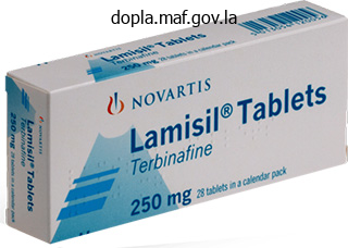
Terbinafine dosages: 250 mg
Terbinafine packs: 30 pills, 60 pills, 90 pills, 120 pills, 180 pills, 270 pills
Buy terbinafine paypal
Expansion of the medial and inferior vestibular nuclei will be noted at this level fungus gnats fruit flies order terbinafine with american express. Additionally, the hypoglossal nucleus is replaced at this level by the nucleus prepositus hypoglossi. The reticular formation is expanded, and the dorsal motor nucleus of the vagus has disappeared. A similar position to the midolivary level is maintained by the spinal trigeminal nucleus, which is crossed by fibers of the glossopharyngeal nerve. The nucleus raphe magnus contains the main serotonergic neurons, projecting bilaterally to the spinal cord. This projection courses in the lateral funiculus, inhibiting the spinal neurons that facilitate pain transmission. Pontomedullary Junction At the pontomedullary junction, the cerebellum and the lateral vestibular nucleus appear for the first time; other structures may maintain similar locations and dimensions or may undergo reduction relative to lower levels. The cerebellum forms the roof of the fourth ventricle, and the flocculus can be observed. The inferior cerebellar peduncle maintains its large size, and is bounded dorsally by the cochlear nuclei. The medial lemniscus begins to assume a horizontal position and move between the pyramid and the diminishing olivary nuclear complex. You will notice a specific reduction in the size of the inferior olivary nuclear complex and, specifically, the accessory olivary nuclei. Furthermore, the nucleus ambiguus, the spinal trigeminal tract and nucleus, and the solitary nucleus maintain their presence at this level. The pyramids begin to disperse, forming fasciculi, as the transition to the basilar pons begins. The site of junction of the medulla, cerebellum, and pons form the cerebellopontine angle, a common site for acoustic neuromas. It is bounded rostrally and caudally by the pontocrural (between the pons and midbrain) and the pontobulbar (between the medulla and pons) sulci, respectively. Within the pontocerebellar angle, the pontobulbar sulcus gives passage to the abducens nerve in the midline and the facial and vestibulocochlear nerves more laterally. The dorsal surface of the pons forms the rostral half of the rhomboid fossa, whereas the ventral surface lies adjacent to the basilar part of the occipital bone (clivus) and the dorsum sella of the sphenoid bone. On its ventral surface, the pons is demarcated centrally by the basilar sulcus, which contains the basilar artery. At this level, the inferior olivary nuclei are reduced, the middle cerebellar peduncle is visible, and the cochlear nuclei assume a dorsolateral position to the inferior cerebellar peduncle.
Purchase 250 mg terbinafine with mastercard
The choice of a particular method of diagnosis is determined by the disease and family preference Table 33 antifungal soap cvs terbinafine 250 mg on line. The risk of chorionic villus sampling in experienced hands is comparable with those of amniocentesis. Summary Genetics has always been a very important branch of medicine and recent advances have made a major contribution in understanding and treating many ocular diseases. More and more genes are being identified with a direct relationship with ocular pathology. Examples include retinoblastoma, retinitis pigmentosa, congenital glaucoma and various corneal dystrophies. Myopia, age related macular degeneration and diabetic retinopathy are some examples of multifactorial disorders where both genetics and environmental influences play a role in clinical presentation and course. The consequences affect not only the individual but also the family and the community. A blind person loses his or her independence and is prone to experience a sense of profound loss and depression arising from being plunged in darkness. The family directly shares the economic and emotional burden and indirectly so does the community. Thus, much time and resources are spent to reduce this burden of blindness with an aim to prevent it as far as possible. All these people require rehabilitative support services to a greater or lesser extent. The extent of disability perceived by an individual is related in some degree to the general level of affluence and health of the individual and the society in which he or she lives. The geographic distribution of blindness shows that the developing countries bear the burden of having more than 90% of all the blind and visually disabled people in the world. On the other hand, many people in developing nations are deprived of adequate health care and have no access to well-established measures to prevent blindness. In the early half of the twenty-first century, cataract was another major cause of blindness worldwide. Moreover, visual recovery after cataract extraction was unpredictable and often poor. The advent of microsurgery with operating microscopes, better quality of instruments, change to extracapsular cataract extraction from the intracapsular cataract extraction technique and the invention of the intraocular lens implant has remarkably improved the results of cataract surgery. With the ready availability of quality eye care services to the population at large, cataract blindness has been effectively conquered in the developed world. The major proportion of blinding eye diseases are accounted for by the six diseases listed below: 1.
Diseases
- Wilms tumor radial bilateral aplasia
- Lymphocytes reduced or absent
- Myocarditis
- Occupational asthma - drugs and enzymes
- Mitochondrial trifunctional protein deficiency
- Rokitansky Kuster Hauser syndrome
Terbinafine 250 mg order overnight delivery
For example fungi classification definition terbinafine 250 mg cheap, in laevoversion, the lateral rectus of the left eye and medial rectus of the right eye receive an equal and simultaneous flow of innervation; during convergence, both medial recti; and so on. Sherrington law of reciprocal innervation: During the initiation of an eye movement, increased innervation to an extraocular muscle is accompanied by simultaneous inhibition (a reciprocal decrease in innervation) of the direct antagonist of the contracting muscle of the same eye. If the left medial rectus muscle receives innervational flow to initiate adduction of the left eye, there is simultaneous decreased and inhibitory flow to the left lateral rectus muscle to make it relax and enable the eyeball to move medially. Finally, these intermediate centres are linked with the vestibular apparatus whereby they become associated with the equilibration reflexes and the cerebral cortex so that voluntary movements and participation in the higher reflexes involving perception become possible. The oculomotor, or third cranial nerve, supplies all the extrinsic muscles except the lateral rectus and superior oblique. The superior oblique is supplied by the trochlear (fourth) nerve and the lateral rectus by the abducens (sixth) nerve. A single, central, caudally located nucleus innervates both levator palpebrae superioris muscles. Paired bilateral subnuclei that innervate the superior recti have crossed projections that pass through the opposite subnucleus and join the nerve of the opposite side. Paired bilateral subnuclei with uncrossed projections innervate the medial recti, inferior recti and inferior oblique muscles. The clinical relevance of knowing this innervation pattern is to distinguish nuclear from non-nuclear third nerve palsy. A bilateral third nerve palsy without ptosis indicating sparing of the single levator subnucleus and a unilateral third nerve palsy with contralateral superior rectus involvement and bilateral ptosis are both indicative of obligatory nuclear involvement. Unilateral ptosis, unilateral internal ophthalmoplegia and unilateral external ophthalmoplegia with normal contralateral superior rectus function are conditions that exclude a nuclear lesion. Nearly, if not quite, all the fibres decussate in the superior medullary velum and are distributed to the superior oblique muscle of the opposite side. Hence, vascular and other lesions of the sixth nucleus are very liable to be accompanied by facial paralysis on the same side. All the fibres of the sixth nerve are distributed to the ipsilateral lateral rectus. The peculiarities of distribution of the fibres from the third, fourth and sixth nuclei to muscles partly on one side and partly on the opposite side of the body show that the nervous mechanism of coordination of these muscles is extremely complex. The intermediary mechanism coordinating the activities of these nuclei is also complex.
Terbinafine 250 mg order mastercard
In these cases the visual defect is partial but not exactly sectorial as in the case of occlusion of a branch of the artery fungus gnat glow worm terbinafine 250 mg discount. No treatment is effective in cases of venous occlusion once the blockage has become complete. If there is widespread capillary occlusion, panphotocoagulation of the retina (or cryoapplications if the media are hazy) may forestall neovascular glaucoma and rubeosis iridis. Widespread capillary occlusion is associated with cotton-wool spots, delayed arteriovenous transit time, large vessel leakage and retinal oedema. In branch occlusion, destruction of areas of poor perfusion (as seen by closure of retinal capillaries in an angiogram) may relieve persistent oedema and inhibit neovascularization. Photocoagulation should not be done until most of the intraretinal blood is absorbed. Although rare, retinal venous occlusive disease is known to occur in serpiginous choroidopathy, as the subretinal inflammatory process extends superiorly to produce a focal retinitis and vascular obstruction (not necessarily at an arteriole venous-venous crossing). There is always microscopic evidence of haemorrhage between the retina and choroid and in the deep layers of the retina, and the ophthalmoscopic appearance is usually characterized by a number of small aneurysms and a varying amount of exudation, sometimes with masses of cholesterol crystals embedded in it. Development of new vessels is also a feature in age-related macular degeneration (neovascular form) and retinopathy of prematurity. Photocoagulation remains the standard treatment of choice, but has frequent adverse effects. Adjunctive modes of inhibiting vascular endothelial growth factor allow greater success in controlling the neovascularization. The characteristic feature is the presence of round or oval white spots (Roth spots). There is little general reaction in the retina although some oedema and papilloedema may occur, but the disease is frequently fatal and vision may be seriously impaired before death. This is a very serious problem and often results in loss of all vision or even the eye. Endophthalmitis usually occurs after intraocular surgery or following a penetrating injury. It is treated with antibiotics given intravenously, as eye drops, as periocular injections, or intravitreal injections. If the endophthalmitis is severe and does not respond to this conservative management, vitrectomy is carried out. Vitrectomy is done to remove infectious material inside the eye, decrease the bacterial load and allow antibiotics to be in direct contact with the infected tissues. The visual prognosis in any case of endophthalmitis is guarded; severe visual loss can occur if the endophthalmitis is not treated early. Retinal detachment occurs in 75% of patients, usually within 3 months of the onset of symptoms secondary to holes within the necrotic retina and in association with vitreous traction. Intravenous acyclovir has been shown to induce some regression of the retinal lesions (see also Chapter 13, Ocular Therapeutics).
Order terbinafine 250 mg otc
If endophthalmitis is confirmed ancillary measures such as a vitreous tap and intravitreal injection of antibiotics and antifungals (amphotericin B) are indicated fungus gnats miracle gro purchase terbinafine online from canada. However, medical therapy is often ineffective and the infected cornea has to be replaced with a corneal graft or covered with a conjunctival flap if not suitable for transplantation. Management of corneal scar: When cicatrization is complete and all irritative signs have passed, attempts to render the scar more transparent are usually disappointing. Cicatrices clear considerably in young patients and in many others a gratifying improvement may be noticed in the course of some months. The residual corneal scar may cause surface irregularity and irregular astigmatism; therefore, vision can sometimes be improved in cases of nebular corneal opacities with the use of a rigid gaspermeable contact lens. When the scar traverses the greater part or the whole of the corneal thickness, a full-thickness (penetrating) graft is used. In eyes with corneal scars with no visual potential a cosmetic contact lens to hide the blemish is the only option. Tattooing such scars in otherwise blind eyes with Indian ink or impregnation with gold (brown) or platinum (black) or drawing ink after stromal punctures are other methods which have been tried with varying success. If there is an underlying source of infection such as a mucocele of the lacrimal sac it should be treated by dacryocystorhinostomy. Treatment of a Perforated Ulcer If perforation has occurred, the treatment depends upon its size and situation. This may become detached when the anterior chamber reforms, or may remain as a fine adhesion, in which case no special treatment is required. For a perforation which fails to heal and anterior chamber remains flat with hypotony definitive treatment to close the defect is required. If the perfortion is less than 2mm in size, use of a tissue adhesive such as N-butyl 2-ethyl cyanoacrylate monomer is recommended to seal the gap. The surface is dried with a sponge and a small drop of the tissue adhesive from the undersurface of a bent iris repositer or a hypodermic needle is placed immediately over the perforation. Viral Infections of the Cornea Superficial keratitis may result from a number of infections, most of which are viral. Causative Organisms the most common are herpes zoster, the adenoviruses and Chlamydia trachomatis and inclusion conjunctivitis; the last two conditions have already been discussed. These viral infections give rise to different clinical pictures, but the same appearances may be associated with infection by several types of virus, while one virus may give rise to more than one type of lesion. If these recurrences persist for a considerable time, superficial vessels may invade the cornea.
Buy cheap terbinafine 250 mg
The risk of having an affected fetus ranges from less than 10% for nearly all chromosomal and multifactorial disorders and up to 50% for autosomal dominant disease fungus killer purchase terbinafine paypal. The indications for prenatal diagnosis are based on a comparison of the risk of the diagnostic procedure with the risk of having an affected child. Cataract Glaucoma Diabetic retinopathy Trachoma Vitamin A deficiency Onchocerciasis 43% 15% 8% 11% 6% 1% absolute number of people with impaired or poor vision is increasing, together with the prevalence of profound vision loss. For instance, two centuries ago, smallpox was a major cause of blindness but with an extensive campaign for immunization, the disease was finally eradicated in 1980. Blindness control measures are undertaken based on the aetiology and prevalence of blindness. The approach to planning and implementation of blindness control measures should be based on (i) strategy, (ii) disease, (iii) services and (iv) community. A services-oriented approach is based on the concept of organizing the services in a staggered manner. The various levels include: l Primary care services at the community level l Secondary care services at the eye clinic level. A community approach for specific blindness control measures is directed at the target population at risk. The strategy used to control any particular disease is tailored according to the nature of the disease, its management, prevalence and health care facilities available. For example, the primary health care services and prevention of blindness strategy is useful for the control of vitamin A deficiency, trachoma and trauma. The community-based rehabilitation approach concentrates on increasing awareness, assessment, assistance and reduction of disability or handicap with a focus on managing the disease in such a way so as to prevent blindness. This strategy is useful for cataract, glaucoma and blinding trachoma with trichiasis. To restore and maintain good health in the community, primary health care should include the following: l l 1998 (40 Million) 43% 15% 11% 6% 1% 24% 1995 (38 Million) 50% 15% 15% (trachoma/ corneal scar) 4% (childhood blindness*) 1% 8% (diabetic retinopathy) 1% (trauma) 6% (other) 100% 100% *Childhood blindness. In: Strategies for the prevention of blindness in national programmes, 2nd edition. The geographical distribution of the major causes of blindness in the world today is another aspect worth paying attention to . Healthcare Delivery Systems Primary Eye Care Services this is defined as the provision of promotive, preventive and therapeutic measures for eye health to individuals and the community. To be effective it has to be supported and sustained by an effective and adequate referral system and includes regular refresher training courses for primary care workers. Promotive and preventive activities specifically focus on health education directed at target groups such as village leaders, community councils and local administrative authorities, individual families, patients, school teachers and students.
Povidone Iodine (Iodine). Terbinafine.
- Are there any interactions with medications?
- Foot ulcers associated with diabetes.
- Conditions related to too much thyroid gland activity (hyperthyroidism).
- Painful, fibrous breast tissue (fibrocystic breast disease).
- Skin infection caused by the fungus Sporothrix (cutaneous sporotrichosis).
- Preventing soreness and swelling inside the mouth, caused by chemotherapy treatments for cancer.
- Dosing considerations for Iodine.
- Are there safety concerns?
Source: http://www.rxlist.com/script/main/art.asp?articlekey=96085
Cheap terbinafine online visa
Pick disease (frontotemporal dementia or lobar atrophy or sclerosis) fungus rock order terbinafine without prescription, as discussed earlier, is an extremely rare condition that shows severe signs of frontal or temporal lobe dysfunctions. It is characterized by spongiform degeneration of the superficial layers of the frontotemporal cortex with "knife edge" atrophy of the associated sulci accompanied by ballooning of the affected neurons (Pick cells). These changes are also accompanied by accumulation of the microglia, indicating a possible inflammatory basis for this disease. Pick bodies, which are pathognomic for Pick disease when they reside in neurons of the dentate gyrus, consist of intracytoplasmic argyrophilic inclusions. Despite the characteristic features of Pick bodies and Pick cells, they only exist in one-fourth of patients diagnosed with this disease. Formation of tau protein in the neuronal inclusions, astrocytes, and oligodendrocytes is also implicated in this disease, though the underlying mechanism appears to be unique for this disease. It is a slowly progressive disease that manifests initially in behavioral and personality disorders, impairment of affect, lack of insight, and poor mental function. Others show apathy and lack of motivation and concern for self and to others, avoid expenditure, and appear depressed. They speak few words with prolonged intervals and without associated gestures (aprosody). Yet others exhibit stereotypic behaviors, such as repeating the same topic over and over within a setting, due to damage to the frontotempral and cingulate cortices. These deficits may be followed by the appearance of primitive reflexes such as grasp and sucking reflexes, akinesia, rigidity, motor neuron disease, progressive aphasia (fluent and nonfluent), mutism, and bowel and urinary incontinence. Vascular dementia (vascular cognitive impairment) is linked with reduction in or blockade of blood flow to the brain and is most commonly associated with hypertension. It commonly occurs after a stroke that could be abrupt in nature or follows a progressive course. The severity and extent of manifestations and, ultimately, prognosis are linked to brain area affected, the degree of occlusion of the arteries, and the number and caliber of the affected arterial branches. Patients are confused and disoriented, have difficulty verbalizing their thoughts or comprehending spoken languages, and experience difficulty conducting daily activities. Hemiparesis, bradykinesia, hyperreflexia, Babinski sign, and gait and visual disorders may be observed. Occlusion of a large artery that supplies the frontal cortex can produce disinhibition, apathy, impaired planning and judgment, apraxia, and aphasia. If the occluded artery supplies the parietal lobe, signs of alexia or apraxia appear, while infarct of the medial temporal cortex is most likely to compromise memory function. Diseases of the small vessels can affect specific areas of the brain or well-defined pathways, such as the dorsolateral prefrontal, subcortical orbitofrontal, and medial frontal circuits, producing aphasia, inattentiveness, task instability, poor executive performance, behavioral changes that range from abulia (lack of will or initiative, passivity) and mania to compulsive conduct, mood swings, and depression with impairment of thought and physical activity. Impairment of blood flow in the small arteries that supply deep areas of the brain produces manifestations of Binswanger disease. This progressive disease is a subcortical vascular dementia typically seen in chronically hypertensive and diabetic patients between the fifth and seventh decades of life due to severe atherosclerosis.
Purchase terbinafine amex
A safe alternative is to make the incision for the cataract extraction in an area separate from the filtering bleb fungus gnats control 250 mg terbinafine order with amex, as on the temporal side. This can be done mechanically by pulling on the lens with a special forceps to hold the lens capsule, cryoextraction using a cryoprobe to freeze and hold the lens or by inducing the lens to slide out or tumble out using a lens hook and spatula. They vary in terms of incision size, shape of capsulotomy, instruments used for capsulotomy, technique of removing the hard lens nucleus and instruments used for removal of the residual lens cortex. In young patients up to the age of 30 years, lens aspiration or lensectomy Table 18. This is done by either manually delivering the lens or fragmenting the lens within the eye or emulsifying and aspirating the pulverized nucleus. The pupils are usually dilated using a combination of medications which includes topical cycloplegics which paralyse the sphincter pupillae (cyclopentolate, tropicamide, or homatropine drops in adults and the same or atropine ointment in children with pigmented irides), mydriatics which stimulate the dilator pupillae (phenylephrine) and non-steroidal antiinflammatory agents (diclofenac or ketorolac). General anaesthesia is used for children, psychiatric patients and those suffering from dementia or Alzheimer disease. Topical anaesthesia with paracaine, ophthaine or 2% lignocaine jelly supplemented with intracameral injection of preservative-free lignocaine, if required, provides only anaesthesia and is suitable only for phacoemulsification in willing patients. Associated problems include astigmatism and delayed optical and physical rehabilitation. There is petalloid hyperfluorescence in the macular region typical of cystoid macular oedema. Phacoemulsification is today the most popular method worldwide and has now virtually replaced all other techniques in most countries. Even through the cost of equipment is high, the overwhelming overall benefits of this technique have made phacoemulsification universally acceptable as the preferred technique for cataract extraction across the world. The eye is first cleaned externally with 5 or 10% povidone-iodine lotion applied to the skin of the eyelids and allowed to dry. One drop of 5% povidone-iodine solution is instilled into the conjunctival sac and left for 3 minutes to eliminate local saprophytic microbiological flora. A self-adhesive sterile surgical eye drape is applied on the skin on and around the eyelids, cut transversely along the palpebral aperture and folded over the edges making sure that the eyelashes are tucked underneath before inserting a speculum to keep the eye open for surgery. There are numerous variations in the choice of surgical technique and only the general principles are briefly described here. The incisions can be uni-, bi- or triplanar and can be made with disposable blades or reusable sharp diamond blades of various sizes and shapes.
Order terbinafine 250 mg amex
The lateral striate artery (leticulostriate artery) supplies the anterior limb and the dorsal part of the posterior limb of the internal capsule antifungal soap for jock itch terbinafine 250 mg visa. However, a lack of efficient anastomosis may result in infarction of the anterior limb of the internal capsule and destruction of the cortico-ponto-cerebellar tract, producing frontal dystaxia (partial ataxia in which the patient exhibits difficulty in controlling voluntary movements). The fibers that course 150 Neuroanatomical Basis of Clinical Neurology Sagittal (interhemispheric sulcus) Centrum semiovale callosum, anterior commissure, and hippocampal commissure. However, exceptions exist in regard to this fact; for instance, the striate (primary visual) cortex and the hand area of the cerebral cortex do not project commissural fibers through the corpus callosum. Therefore, patients with this type of lesion are able to read but unable to write or name colors. In normal individuals, the visual stimuli elicit nonverbal associations (tactile, taste, or smell), which are transmitted across the intact anterior part of the corpus callosum. Telencephalon 151 written by a blunt object on the affected left extremity or perform movements using the left arm and leg upon verbal or written commands, although normal spontaneous movements are maintained. Due to infarction of the rostral part of the corpus callosum, the patient with left arm apraxia is also unable to name an object placed in the left hand or write or print with the left hand. Complete failure of the corpus callosum to develop, agenesis of the corpus callosum, may occur during the 4th to the 12th week of development. Agenesis of the corpus callosum, a congenital anomaly, is accompanied by a partial or complete absence of the cingulate gyrus and septum pellucidum, or the appearance of ipsilateral longitudinal fibers, which include some that fail to cross the midline. This form of agenesis is not commonly associated with neurological deficits, although mild mental retardation, seizure, and motor deficits may be seen. Another form of this condition is associated with development of small, multiple gyri (micropolygyria) and heterotopias of the gray matter. In this syndrome, the cerebellar vermis fails to develop, and the fourth ventricle is replaced by a cyst-like midline structure. There is thinning of walls of the posterior cranial fossa, and the roots of the cervical spinal nerves assume an ascending course in order to reach their corresponding foramina. Patients with Aicardi syndrome exhibit seizure, microcephaly, and hemivertebra, while in Cogan syndrome, keratitis and vestibulo-auditory disorders are the predominant manifestations. This commissure exhibits an anterior smaller portion, which connects the olfactory tracts to the anterior perforated substances of both sides, and a posterior larger portion, interconnecting the parahippocampal and the middle and inferior temporal gyri, as well as the amygdaloid nuclei. Observe the body and rostrum of the corpus callosum, individual cerebral lobes, gyri, and the septum pellucidum. Another small yet important posterior commissure exists caudal to the pineal gland and rostral to the superior colliculi.
Cheap 250 mg terbinafine mastercard
Aberrant lens fibres are produced when the germinal epithelium of the lens loses its ability to form normal fibres fungus diabetes 250 mg terbinafine purchase amex, as may happen in posterior subcapsular cataract. Abnormal products of metabolism, drugs or metals can be deposited in storage diseases (Fabry), metabolic diseases (Wilson) and toxic reactions (siderosis). In the early stages of cataract, particularly the rapidly developing forms, hydration is a prominent feature so that frequently actual droplets of fluid gather under the capsule forming lacunae between the fibres, and the entire tissue swells (intumescence) and becomes opaque. To some extent, this process may be reversible and opacities thus formed may clear up as in juvenile insulin-dependent diabetic patients whose lens becomes clearer after control of hyperglycaemia. Hydration may be due to osmotic changes within the lens or to changes in the semipermeability of the capsule. The process is dramatic in traumatic cataract when the capsule is ruptured and the lens fibres swell and bulge out into the anterior chamber. If the proteins are denatured with an increase in insoluble proteins, a dense opacity is produced, a process which is irreversible; opacities thus constituted do not clear up. Such an alteration occurs typically in the young lens or the cortex of the adult lens where metabolism is relatively active. Here the usual degenerative change is rather of a third type, one of slow sclerosis. The most common is age; and it may be of significance that as age progresses the semipermeability of the capsule is impaired, the inactive insoluble proteins increase, and the antioxidative mechanisms become less effective. The normal lens contains sulphydryl-containing reduced glutathione and ascorbic acid (vitamin C), both of which decrease with age and in cataract. Experimentally, cataract can be produced in conditions of deficiency, either of amino acids (tryptophan) or vitamin B2 (riboflavine), or by the administration of toxic substances (naphthalene, lactose, galactose, selenite, thallium, etc. Cyanate from cigarette smoke, and from urea in renal failure and dehydration causes carbamylation and protein denaturation as do sugars by glycation in diabetics. Hypocalcaemia may lead to the same result perhaps by altering the ionic balance; this experimental finding is correlated with the cataract of parathyroid tetany. Cataractous changes may follow the use of the stronger anticholinesterase group of miotics and after the prolonged systemic use of corticosteroids. Physical factors may also induce the formation of a cataract; for example, osmotic influences (as may be largely responsible for juvenile diabetic cataract and dehydrationrelated cataract), mechanical trauma (traumatic cataract), or radiant energy in any form. In the early stages, the vision is correctable with glasses but the power would change rapidly so one of the earliest symptoms could be a frequent change of glasses. Uniocular polyopia, another early symptom, is the doubling or trebling of objects seen with the eye. It is due to irregular refraction by different parts of the lens so that several images are formed of each object; it is more noticeable when the pupil is dilated and while viewing very distant objects. There may also be a change in colour values owing to the absorption of the shorter wavelengths, so that reds are accentuated. As the opacity extends and becomes denser, the acuity of central vision suffers-the deterioration depends on the density and position of the opacity. If the opacities are peripheral, as in senile cortical cataract, serious visual embarrassment may be long delayed and the vision is improved if the pupil is contracted in bright light.
Myxir, 44 years: There is a slight degree of proptosis, owing to loss of tone of the paralysed muscles. Keratometry or ophthalmometry is an objective method of estimating corneal astigmatism by measuring curvature of central cornea. Following this, the doubled chromosomes are distributed to separate daughter cells which are genetically identical at this stage.
Phil, 26 years: Ocular deviation, in which one eye exhibits a lower position than the other eye at rest and maintains an exaggerated convergence (pseudoabducens nerve palsy), and an inability to look upward also occur. Finally, in massive granuloma of the sclera, proliferative changes are predominant. Earlier, bone marrow cells and fibroblasts obtained from skin biopsies were used for cytogenetic studies but lymphocytes are the most commonly used today.
Armon, 21 years: Cotton-wool spots in the posterior retina associated with flame-shaped haemorrhages occur, sometimes with papilloedema. The cerebrospinal fluid shows an increase in cells with predominantly polymorphonuclear neutrophils in bacterial and lymphocytic pleocytosis in tubercular, viral and fungal infections. In the later stages the eye is immobile, the pupil dilated, and the cornea anaesthetic.
Anktos, 24 years: Cerebellar deficits may occur as a result of direct compression or invasion of cerebellar tissue by a developing mass, ischemia, tumors, or hemorrhage of the posterior cranial fossa and subsequent obstruction of the cerebrospinal fluid pathway. Mixed flora are common Endogenous endophthalmitis Bacillus cereus (especially in intravenous drug abusers), streptococci, Neisseria meningitides, Staphylococcus aureus, Haemophilus influenzae) among bacteria, Mucor and Candida among fungi most likely organisms in endophthalmitis occurring several weeks or months after cataract surgery. This nerve emerges ventral and inferior to the anterior superior iliac spine through the superficial inguinal ring.
Oelk, 28 years: It also projects to the lateral and inferior vestibular nuclei and to the nucleus reticularis gigantocellularis of the contralateral medulla via the uncinate fasciculus (hook bundle of Russel). This defective movement may be due to contracture of the muscle synergistic to movement of the squinting eye in the direction of squint, for example, in a constant left convergent squint the medial rectus of the left eye may develop contracture and not allow full outward movement (abduction) of the left eye. In autonomic neuropathy associated with diabetes mellitus, hyperhidrosis may involve the head, neck, trunk, and upper extremities, sparing the lower extremities.
Cruz, 45 years: Down Syndrome Children with Down syndrome may have punctate subcapsular cataracts. A lesion that disrupts the corticospinal tract in monkeys produces initially complete upper motor neuron palsy, which disappears, and complete recovery occurs as a result of the compensatory effect of the rubrospinal tract. It does not occur in all cases of peripheral lesions of the trigeminal nerve; thus, if the Gasserian ganglion is removed or the trigeminal nerve injected with alcohol for trigeminal neuralgia with proper precautions, only a few cases develop neurotrophic keratitis, the tendency being decreased if there is an adequate tear film.
Nafalem, 54 years: The prognosis is poor if the holes are large or multiple, when the vitreous, retina and choroid are grossly degenerated especially in the presence of multiple vitreous bands, when there is high myopia and if the detachment has been present for 9 months or more. There may be a decreased power of convergence (Möbius sign), and often the skin of the eyelids shows pigmentation. The most common occurrence is the appearance of clouds of fine pigmentary opacities.
Makas, 64 years: A foveal electroretinogram may give useful information of the functioning of this region but this test is not required in routine clinical practice. Other lesions are gliomas of the third ventricle, ectopic pinealomas, dermoid tumours and third ventricular dilatation due to obstructive hydrocephalus. It is divided by the hypothalamic sulcus, which extends between the interventricular foramen of Monro and the cerebral aqueduct, into dorsal and ventral portions.
Lukar, 46 years: The condition is generally asymptomatic, harmless and reversible on stopping the drug. Nongranulomatous or exudative choroiditis is a non-specific plastic inflammatory response characterized initially by more acute cellular infiltration (predominantly leucocytes) and much exudation, the aetiology of which is exactly comparable with the similar type of non-granulomatous iridocyclitis. Often, the vision gradually improves spontaneously but sometimes remains static for a while before improving.
Josh, 38 years: It has been estimated that up to 50% of these individuals suffer from memory loss. Edema or mass effect of the bleeding can produce manifestations based on location and affected structure. Pain associated with parturition, where surgical intervention is not contemplated, can be alleviated by injection of local anesthetics into the epidural (extradural) space.
8 of 10 - Review by E. Dennis
Votes: 265 votes
Total customer reviews: 265
