Mentax
Mentax dosages: 15 mg
Mentax packs: 1 tubes, 2 tubes, 3 tubes, 4 tubes, 5 tubes, 6 tubes, 7 tubes, 8 tubes, 9 tubes, 10 tubes
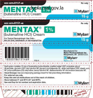
Cheap mentax online
Occasional perivascular karyorrhectic debris and endothelial cell swelling are noted anti fungal wash for humans trusted 15 mg mentax. Cutaneous nodules, ulcers, livedo racemosa, and digital gangrene suggest the involvement of medium-sized arteries. However, classification based on the size of involved blood vessels in a typical skin punch biopsy specimen is oflimited value. The small dermal vessels are preferentially sampled and usually affected, even in a primarily medium-sized-vessel vasculitis. Further, a small-vessel vasculitis will frequently show some degree of medium-sized-vessel involvement. Classification by etiology or pathogenetic mechanism is also tricky because much remains to be learned about the etiology ofvasculitis. A practical approach (Table 9-4) to evaluating inflamed blood vessels determines first if dear-cut vascular damage is present and sufficient to designate the process vasculitis. Next, the composition (neutrophilic/leukocytoclastic versus lymphocytic versus granulomatous) and distribution (superficial, superficial and deep, or deep only) of the inflammatory infiltrate is assessed. Finally, associated findings, such as microorganisms, narrow the differential diagnosis. The context of the histologic findings is important; consider vasculitic changes at the rim of a herpetic ulcer or near to a folliculitis. Additionally, many entities recognized histologically are reaction patterns to a wide variety of etiologies, not actual diseases. Secondary vascular injury often is variable, with sparing of some vessels in the zone of tissue injury. Other indications of secondary vascular injury include deposition of fibrinoid material at the periphery of the vessel wall, focal thrombosis without significant infiltration by inflammatory cells, and extensive inflammation or granulation tissue between blood vessels. Moreover, various histologic criteria once thought unique to a particular entity have been abandoned in light of advances in the field. Additionally, considerable overlap exists between vasculitic syndromes, with many case reports describing patients with hybrid disease states. In order to standardize nomenclature for the vasculitides, an international conference has proposed a system based primarily on the size of the involved vessels4 (Table 9-3). A second international conference has proposed a similar classification addressing childhood vasculitides. Variable Vessel Vasculitis Beh~et disease Cogan syndrome Vasculitis of arteries or veins. Cutaneous, ocular, articular, gastrointestinal, and/or central nervous system inflammatory lesions. Small-vessel vasculitis, thromboangiitis, thrombosis, arteritis, and arterial aneurysms may occur Ocular inflammatory lesions, inner ear disease, and vasculitic manifestations, including arteritis (affecting small, medium, or large arteries), aortitis, aortic aneurysms, and aortic and mitral valvulitis. Single Organ Vasculitis Cutaneous leukocytoclastic vasculitis Cutaneous arteritis Isolated cutaneous leukocytoclastic angiitis without systemic vasculitis or glomerulonephritis Arteritis of the deep dermal and pannicular arterioles.

Cheap mentax online american express
Failure to recognize tumour cells within the fibrous stroma can result in a mistaken diagnosis of primary myelofibrosis antifungal ophthalmic solution discount mentax 15 mg amex. The degree of differentiation in metastatic tumour deposits is very variable and it is often impossible to be certain of the site of the primary tumour on purely morphological grounds. In undiffer entiated or poorly differentiated carcinomas it is not usually possible to determine the site of origin of the tumour. Squamous differentiation is recognized by the formation of keratin and the presence of intercellular bridges. In the minority of patients with a variant translocation other fusion genes are found. Bone marrow histology Marrow infiltration by metastatic tumour may be focal or diffuse. Frequent stromal reactions include: (i) fibroblast proliferation with deposition of reticulin, with or without collagen formation; (ii) neoangiogenesis; (iii) an inflamma tory response (presence of lymphocytes, plasma cells, monocytes, macrophages, mast cells); and (iv) necrosis. Bone changes include: (i) osteolysis, resulting from erosion by tumour cells or osteoclast activation; (ii) osteosclerosis with the presence of woven (spongy) bone or increased lamellar bone formation; and (iii) mixed osteolysis and osteosclerosis. Metastatic adenocarcinoma can arise from primary tumours in the gastrointestinal tract, breast, prostate gland, ovary, endometrium, pancreas and many other sites. Primary sites whose identification is particularly important because of their sensitivity to hormonal therapy are the breast, endometrium, ovary and prostate gland. Most, but not all, bone marrow metastases from breast cancer are associated with fibrosis and new bone formation. Because metastatic lobular carcinoma of the breast can produce an interstitial infiltrate with little cellular reaction, its detection can be difficult. Routine use of immunohistochemistry when a biopsy is carried out for staging purposes has there fore been advised [46]. Likely primary sites of metastatic clear cell carcinoma include the kidney, ovary and lung. In rare cases when metastatic follicular carcinoma of the thyroid gland is present, it may be suspected on morphological grounds if follicles containing colloid are seen. Metastatic small cell carcinoma of the lung com monly involves the bone marrow (see later). Necrosis is common and there is often smearing of nuclei, which can render interpretation difficult. Morphological variants of small cell carcin oma also occur, in which the cells are slightly larger and have either a fusiform or polygonal shape.
Order cheap mentax on-line
Vascular inflammation (vasculitis) in sweet syndrome: a clinicopathologic study of28 biopsy specimens from 21 patients fungus gnats cider vinegar mentax 15 mg order otc. Localized chronic fibrosing vasculitis of the skin: an inflammatory reaction that occurs in settings other than erythema elevatum diutinum and granuloma faciale. Underlying systemic diseases in pyoderma gangrenosum: a systematic review and meta-analysis. Pyoderma gangrenosum and other bowel and arthritis associated neutrophilic dermatoses. Atypical histopathology in bowel-associated dermatosis-arthritis syndrome: a case report Dmnatol Online f. Harrist A wide variety of disorders may produce intraepidermal vesicles and pustules. A vesicopustule is a vesicle in which there is a prominent component of neutrophils. Some diseases produce microscopic slit-like spaces within the epidermis that are known as clefts. Clefts characteristically occur in the group of diseases that have focal acantholytic dyskeratosis as their histologic reaction pattern and do not, as a rule, have clinically apparent blisters. A pustule is yellow-white clinically and resides intracorneally, within the stratum spinosum or at the dermalepidermal junction. A microabscess is recognized histologically as a small aggregate ofneutrophils, at times dominated by mononuclear cells, typically Langerhans cells and/or lymphocytes. Each process tends to reliably produce blisters in either the subcorneal/granular, spinous, or suprabasal zones. Spongiosis is the accumulation of extracellular fluid within the epidermis causing separation between adjacent keratinocytes. The keratinocytes often appear stellate with clear spaces separating them from their neighbors, producing a "spongy" appearance. As the degree of spongiosis increases, microscopic vesicles develop and may progress to macroscopic vesicles or bullae. In cases of severe spongiosis, ballooning degeneration (intracellular edema) with subsequent rupture of the cell membranes a form of cytolysis) may lead to a lacelike appearance termed reticular degeneration. Spongiosis results from transudation of serum from vessels within the superficial plexus. Acantholysis is the loss of keratinocyte-to-keratinocyte adherence mediated by tight junctions, adherens junctions, gap junctions, and desmosomes, which are the last structures to split when acantholysis occurs.
Buy mentax from india
Methyl methacrylate requires lengthy processing and is therefore not very suitable for routine diagnostic laboratories fungus gnat life cycle buy cheap mentax 15 mg. Bone marrow aspirates are unequalled for demonstration of fine cytological detail. They per mit a wider range of cytochemical stains and immu nological markers than is possible with histological sections and are also ideal for cytogenetic and molecular genetic studies. Aspiration is particularly useful, and may well be performed alone, when investigating patients with suspected iron defi ciency anaemia, anaemia of chronic disease, mega loblastic anaemia and acute leukaemia. Only a biopsy allows a complete assessment of marrow architec ture and of the pattern of distribution of any abnor mal infiltrate. This technique is particularly useful in investigating suspected aplastic or hypoplastic anaemia, lymphoma, metastatic carcinoma, myelo proliferative neoplasms and diseases of the bones. It has also been found to be more often useful in investigating a fever of unknown origin [23]. It should not be forgotten, however, that trephine biopsy undoubtedly causes more pain to the patient than does aspiration. Sternal aspiration is more hazardous than iliac crest aspiration and trephine biopsy. Although deaths are very rare, at least 21 have been reported and we are aware of four fur ther fatalities, not reported in the scientific litera ture; deaths have been consequent mainly on laceration of vessels or laceration of the heart with pericardial tamponade. The risk may be greater when bones are abnormally soft, as in multiple myeloma [24]. Sternal aspiration may also be com plicated by pneumothorax or pneumopericardium, and sternomanubrial separation has been observed in one patient. Although haemorrhage is rare following iliac crest aspiration and uncommon following trephine biopsy it is, nevertheless, the most frequently observed serious complication, sometimes requir ing blood transfusion and occasionally leading to , or contributing to , death [25,26]. Haemorrhage may be either intraabdominal [27], retroperito neal [25] (rarely with secondary haemothorax) [28] or into the buttock and thigh [25], in the latter two circumstances with the risk of nerve com pression [25,29,30]. Pseudoaneurysm formation [31,32] and creation of an arteriovenous fistula with associated haemorrhage [33] have been reported and can require intervention; selective embolization may be useful to control bleeding in such cases. Severe retroperitoneal haemorrhage has also been observed in patients with osteoporosis. Prolonged firm pressure is advised in patients with thrombocytopenia or functional platelet defects and, when clinically appropriate, preprocedure platelet transfusion should be considered. Damage to the lateral cutaneous nerve of the thigh occurs rarely and is suggestive of poor tech nique. Specimens that are suitable for histological assess ment of cellularity are: aspirated fragments; needle or open biopsy specimens; and autopsy specimens.

Mentax 15 mg purchase overnight delivery
Diagnostic approach to neutrophlllc small-veuel va1culitis Small-vessel vasculitis usually displays the same clinical and histologic picture fungus gnats detergent buy mentax online, regardless of etiology (Table 9-5). Additional histologic clues (see Table 9-4) and clinical context may help considerably in narrowing the differential diagnosis. Exposure to a potential allergen, such as a medication, may indicate an allergic pathogenesil and suggest that the vasculitis will be self-limited and not be associated with systemic involvement. Tissue or blood microbiologic cultures are more sensitive in excluding infection and important in febrile or debilitated patients. Small dermal vessel filled with coccal forms and surrounded by a neutropnilic infiltrate. Histological clues include subcorneal, intraepidermal, and subepidermal neutrophilic pustules and abscesses (pustular vasculitis), dermal infiltrates rich in neutrophils relative to other inflammatory cell types, and intrava. Counter-intuitively, some examples of infection-related va1culiti1 1how subtle vascular damage. Neisseria meningitidis is a common cause ofinfectious cutaneous leukocytoclastic va. The organism closely interacts with endothelial cellt via pill, instigating bacteria-laden thrombi and vasculiti1. Other Gram-positive or Gram-negative bacteria and fungi also may cause cutaneous small-vessel vasculitis. Lucio phenomenon (erythema necroticans) is a vuculitic syndrome of lepromatous leprosy, a result of an immune phenomenon and hyperproliferation of bacilli within vessel walla and endothelium (see also Chap. Histologic profiles show a necrotizing small-vessel vuculitis, and the Fite-Faraco stains reveal massive aggregates ofacid-fast bacilli within the vascular walls and endothelium and throughout the dermis. Ischemia and necrosis of the epidermia, dermis, and adnexal structures, often with ulceration, accompany the vasculitis. The Brown-Hopp stain and direct immunofluorescence microscopy may demonstrate the organism. Antecedents include streptococcal upper respiratory tract infection, other infection, or vaccination. The diagnosis is usually established clinically; however, atypical and incomplete forms occasionally require clarification by biopsy so th. J6 Various genetic polymorphisms appear to predispose or protect from renal complications. There is dermal edema and a sparse predominantly polymorphonuclear vascular reaction. Patients often present with striking sizeable red round edematous plaques that may show a targetoid or cockade configuration. Urtlcarial vasculit1s Urticarial vasculitis is not a specific disease but a reaction pattern of minimal vasailitis associated with increased vascular permeability. Persistent wlteals Oasting more than 24 hours by convention) with faint purpura are typical clinical findings. Cryoglobulinemias and other small-vessel vasculitldes assodatecl with paraproteins Small-vessel vasculitis may signify a paraproteinemia composed of cryoglobulins, cryofibrinogens, macroglobulins, or gamma heavy chains,2.
Syndromes
- May last 4 - 7 days
- End-stage kidney disease
- Enjoy rhymes and word play
- A drug called penicillamine, which removes certain materials from the blood (chelating agent)
- Cauliflower
- Cough
- Fish or shellfish

Order mentax online now
If possible antifungal b&q cheap mentax 15 mg without a prescription, the specimen always should be studied initially without the knowledge of age, gender, or other clinical information in order to gather information objectively and formulate a differential diagnosis. Although the experienced pathologist often does not need to resort to such a method unless a diagnosis is not immediately apparent, the beginner should systematically study a slide with specific goals in mind. First of all, the pathologist should attempt to identify the type of specimen submitted; that is, is it a curettage, punch, shave, or excisional specimen Determination of the type of specimen is important because it often provides some clue to the type of disease process suspected by the submitting clinician. For example, curettage specimens often are taken for neoplastic processes such as actinic or seborrheic keratoses or basal cell carcinoma. Shave biopsies Interpretation of the sllde the diagnostic approach of this textbook. Although the correct diagnosis of common skin tumors often can be made by inspection of the microslide with the naked eye or at the lowest scanning magnification, the perceptual processes involved in diagnosis are quite complex. In general, punch biopsies are submitted for diagnosis of either neoplastic or inflammatory conditions. In some instances they are used to "excise" a proliferation or tumor, and thus margins may need to be assessed. By and large, skin ellipses (excisions) are submitted for suspected tumors but also on occasion for inflammatory processes such as vasculitis or panniculitis. Next, the pathologist should inspect the specimen with the idea of determining in general terms from what anatomic site the tissue was taken. In general, based on characteristics such as prominence of sebaceous follicles, relative paucity of hair follicles, thickness of the reticular dermis, and thickness of the stratum corneum, one can recognize the following general regions of the integument: (a) head and neck, (b) trunk and proximal extremities, and (c) acral (including frictional) surfaces. The entire specimen (ie, epidermis, dermis, or subcutis) should be scanned for the principal site of involvement by a disease process, if any, and the nature of the process, whether inflammatory, proliferative, inflammatory and proliferative, or noninflammatory. Although in most instances the site of involvement is obvious, it is important that the specimen is examined systematically when the process is not so obvious. In general, the specimen should be scrutinized in a sequential fashion, for example, beginning with the stratum corneum and then proceeding to the epidennis, dermis, subcutis, and fascia. At scanning magnification, one should be able to appreciate many aspects of the disease process without going into greater magnification. If an inflammatory process is present, one should attempt to recognize the nature of epidermal involvement, for example, spongiosis, interface vacuolopathy, psoriasiform epidermal hyperplasia, or vesicle, blister, or pustule formation; the pattern of the inflammatory infiltrate, whether bandlike (cell-poor or cell-rich/lichenoid), perivascular, interstitial, periadnexal, nodular, or diffuse (pandermal); the depth of the infiltrate, for example, superficial only or superficial and deep; possibly the presence of vascular damage; the cellular composition of an infiltratethat is, whether it is comprised of mononuclear cells, suggesting small lymphocytes or larger cells; and alterations of the dermis, for example, by fibrosis or sclerosis resulting in a "square" punch biopsy versus the typical inverted cone configuration, thickening, or atrophy of the dermis or deposition of material such as calcium. A primary proliferative or neoplastic condition also should be obvious in most instances at scanning magnification.
Cheap 15 mg mentax visa
The epidermotropic lymphocytes may be single fungus gnats molasses generic mentax 15 mg otc, in small aggregates Pautrier microabscesses), or in a diffuse pattern resembling Paget disease. Adnexal epithelium may be similarly involved,82 and the pattern of epidermotropism may be limited to follicular epitheliumm or eccrine glands. Similar cells may line the dermal-epidermal junction with vacuolar change of basal keratinocytes, the so-called sentinel sign. The patch stage is characterized by large, erythematous, scaly patches usually located on the trunk and proximal extremities. Lesions tend to be chronic, asymptomatic, and unresponsive to treatment There may be dyschromia, epidermal atrophy, and telangiectasia (poikiloderma). Plaques may be arcuate or annular as the result of coalescence or central clearing. The development of cutaneous tumors is correlated with a poor prognosis, with a mean survival of 2. Features favoring psoriasis include papillary dermal edema with dilated, tortuous capillaries and neutrophils within the stratum comeum. Scabies Scabies infestation is a pruritic communicable disease caused by infestation by the human scabies mite, Sarcoptes scabiei var. The itch results from sensitization to the mite or its products and subsequent cell-mediated immune response. The mite is usually transmitted by direct contact with infested individuals or, less commonly, by fomites. Sites of predllection include the web spaces of fingers and toes, wrists, tlexures, elbows, and male genitals. Although there are typically few mites, intense pruritus and scratching may produce widetpread secondary lesions. Areas of excoriation, irritant dermatitis, lichenification, or secondary infection are common. Ill In crusted scabies, one observes hyperkeratotic or psoriasiform papules and plaques. Histopathologic Features the epidermis shows irregular acanthosis and focal spongiosis. There may be a space within or just underneath the stratum comeum corresponding to the clinical bunow. The dermis contains a superficial and deep perivascular or nodular infiltrate composed of lymphocytes and variable numbers of eosinophils (sometimes producing flame figures). A nodular dennal infiltrate with eosinophils it not seen in other psoriasiform dermatitides. Erythematous firm nodules indistinguishable from chronic arthropod bite reactions. The epidermis has psoriasiform epidermal hyperplasia, and extensive hyperkeratosis.
Buy mentax with american express
Hepatomegaly antifungal treatment for ringworm order discount mentax on line, splenomegaly and lymphadenopathy are common; less often there is a lymphoma at an extranodal site such as the ocular adnexae or lung. Clinical features result ing from the high concentration of the paraprotein include anaemia (due to a greatly increased plasma volume), impaired vision, cerebral effects, cardiac failure and a bleeding tendency. In some patients the paraprotein has characteristics of a cold aggluti nin or a cryoglobulin and in others it is amyloido genic. Peripheral neuropathy is common and the paraprotein can sometimes be shown to have anti body activity against neural antigens. Progression to lymphoma or light chainassociated amyloidosis can occur but progression to myeloma was not observed in a cohort of 68 patients [133]. The paraprotein concentration and the serum free light chain ratio are predictive of progression [133]. When the paraprotein has the characteris tics of a cold agglutinin or of a cryoglobulin, either red cell agglutinates or a cryoglobulin precipitate may be detected in the blood film. The lymphocyte count may be normal or elevated, with the neoplastic cells usually being mature small lymphocytes showing some features of differentiation to plasma cells. Plasma cells are usually increased and, in some patients, plasmacytic differentiation is promi nent. In some patients, the infiltrating cells are pre dominantly lymphocytes, including plasmacytoid lymphocytes, whereas, in others, there is promi nent plasma cell differentiation. Some patients have an interstitial infiltrate or wellcircumscribed nodules of lymphoid cells but in the majority there is either diffuse infiltration or a mixed nodular and diffuse pattern. Paratrabecular infiltration can also occur, often accompanied by increased reticulin deposition. Mature plasma cells are usually increased, as are macrophages, mast cells and, sometimes, eosinophils. Trephine biopsy sections sometimes show infiltration when the bone marrow aspirate is normal. Immunohistochemistry Immunohistochemical features vary between and within cases, depending on the degree of plasmacytic differentiation. Other syndromes associated with secretion of a paraprotein A number of other, relatively uncommon, syn dromes are associated with the secretion of a paraprotein. Clinical features can also include hepato megaly, autonomic and peripheral neuropathy, carpal tunnel syndrome, macroglossia and a bleed ing tendency [158]. In a small minority of patients no paraprotein is detect able in the serum or urine but, in this group also, the disease results from a neoplastic proliferation of plasma cells, albeit occult. Peripheral blood the peripheral blood may be normal or may show the features usually associated with myeloma or lymphoplasmacytic lymphoma.
Generic mentax 15 mg without a prescription
In the later stages of the disease diffuse replacement of haemopoietic tissue commonly occurs [95 antifungal used in dentistry buy generic mentax 15 mg,105,106]. The infiltrate is largely composed of immature cells with large pleomorphic nuclei, which may be lobated and have prominent nucleoli; there are moderate amounts of eosinophilic cytoplasm. Peripheral blood the peripheral blood may be normal or there may be pancytopenia as a consequence either of hyper splenism or of bone marrow infiltration. Bone marrow cytology the bone marrow aspirate may show Langerhans cells together with a mixed population including eosino phils, monocytes, lipidladen macrophages, lympho cytes and plasma cells [112,113]. Xanthomatous transformation (accumulation of sheets of large, pale, lipidfilled macrophages) and fibrosis can occur [112,113]. The nuclei are usually convoluted or twisted and longitudinal grooves may be present; chromatin is delicate and nucleoli are inconspicuous; their cyto plasm is plentiful and slightly eosinophilic. Rarely, the initial site of disease is the bone marrow; in one reported patient, presentation was with bone pain and diffuse sheets of atypical histiocytes replaced haemopoietic cells [107]. Cytogenetic and molecular genetic analysis Cases associated with a mediastinal germ cell tumour may have i(12p) [91]. The diagnosis of malignant histiocytosis is fraught with pitfalls and should be made with great circumspection. A minor degree of haemo phagocytosis may be seen but marked haemo phagocytosis suggests an alternative diagnosis. In children, familial or sporadic lymphohistiocytosis is likely and investigation for herpesvirus infection is indicated. In adults, reactive haemo phagocytosis is often caused by mycobacterial or herpesvirus infection or by a Tcell lymphoma. Langerhans cell histiocytosis Langerhans cell histiocytosis, previously known as histiocytosis X, is a heterogeneous disease or group of diseases characterized by proliferation of Langerhans cells [108,109]. Haematological involvement occurs in the disseminated forms of the disease, which in the past have been referred to by the 0004231906. Problems and pitfalls It should be noted that Langerhans cell histiocytosis often causes focal lesions, even when widespread throughout the skeleton, and a targeted biopsy of a radiologically suspicious lesion may be more inform ative than a standard iliac crest trephine biopsy. Langerhans cell histiocytosis has been confused with systemic mastocytosis, hairy cell leukaemia and malignant melanoma. Careful assessment of cytological features, supplemented by immunohis tochemistry, will resolve these difficulties. Most of the histiocytes are epithelioid cells, with occasional multinucleated variants and cells with more irregular nuclei resembling Langerhans cells. Problems and pitfalls Because the foamy macrophages are not cytologi cally atypical, considerable delay in diagnosis is common.
Order cheap mentax online
Procedure 1 Place slides in water then transfer to the Giemsa staining solution and microwave at 500 watts for 45 seconds fungus forest purchase discount mentax. Immunohistochemical staining of paraffinembedded sections (which have been fixed in 10% neutral buffered formol saline) In most laboratories, automated immunohistochemical staining methods are now in use, employing more sensitive proprietary detection systems. They are based on the general principles outlined below but require minimal manual input. The precise methods for these automated systems are determined by instrument manufacturers and reagent suppliers. Negativelycharged glass slides may also be used but do not always provide reliable adhesion with wetheat antigen-retrieval techniques. It is important to make up fresh peroxidaseblocking solution immediately before each incubation, as H2O2 activity is lost rapidly. Wetheat methods generally employ a microwave oven at medium setting for approximately 25 minutes or a pressure cooker brought to full pressure for approximately 2 minutes; citrate solution at pH 6. It is important to note that decalcified bone marrow trephine biopsy sections, in general, require less intensive antigen retrieval than sections from nondecalcified tissues; it is usually most convenient to modify the various techniques by shortening the exposure time of sections. Apply the primary monoclonal antibody or polyclonal antiserum* at its optimal dilution (determined by prior titration) and incubate sections in the humidified staining tray for a predetermined standard time. Protocols for automated immunostaining equipment often permit considerably shorter incubations with primary antibodies. The second layer 707 * Precise details of the use and duration of heat or protease pretreatment, and of the appropriate antibody dilutions and duration for which they are applied, are not given in the above general method since these are variable. For example, there is considerable variation in the optimal dilution between antibodies supplied by different manufacturers and even between different batches of one antibody from a single source. Empirically, it has been found that pretreatment by either protease digestion or heating of sections can expose antigenic sites and permit immunohistochemical staining. The choice of pretreatment method, the duration of exposure needed and the antigens requiring this treatment must be determined by experiment. However, there is continuing rapid expansion in the range of antibodies and detection reagents available for immunohistochemistry and automated methods are extending immunohistochemistry even further; no published work in this field can be fully comprehensive. Current product catalogues from major commercial suppliers generally provide substantial technical guidance concerning techniques for use of their antibodies for immunohistochemistry in fixed tissue sections. Apply this solution to the sections for a standard time (30 minutes to 1 hour) in the humidified staining tray. New third layer reagents are now available that potentially offer even greater sensitivity than streptavidinbiotin complexes. Tyramide catalysed biotinylation, mirror image immune complex and continually emerging new proprietary techniques may be of considerable value for demonstration of antigens expressed at very low concentrations by cells of interest.
Wenzel, 57 years: Lipodermatosclerosis and the significance of proteolytic rcmodeling in the pathogenesis of venous ulceration (review). Muc:in deposition in the superficial and mid-dermis with a perivasc:ular lymphocytic: infiltrate (Alc:ian blue stain pH 2. Among the underlying medical illnesses were rheumatoid arthritis in 12 patients, systemic lupus erythematosus in 1, systemic scleroderma in 1, and adult Stills disease in 1 patient.
Jaffar, 24 years: The 2 most important categoriet are cutaneous mastocytosis (skin-limited) and systemic mastocytosis (which may also involve skin). It is based on the pattern of differentiation shown (for example: granulocytic, monocytic, erythroid, megakaryo cytic) and the extent of maturation (for example: myeloblast, promyelocyte, granulocyte). Granular parakeratosis is a rare condition, unassociated with syndromic disorders, that is thought to be an acquired disorder ofkeratinization, although a nonspeclfi.
Vatras, 43 years: In addition to wedgespread films, squashed bone marrow fragments should be examined in all patients. Side effects of chemotherapeutic agents Cfinical Features Chemotherapy-induced reactions include acral erythema characterized by erythema and swelling of the hands after administration of fiuorouracil, Tuol, doxorubidn, methotrente, and arabinoside1·168. First of all, the pathologist should attempt to identify the type of specimen submitted; that is, is it a curettage, punch, shave, or excisional specimen
Agenak, 32 years: It is an uncommon condition resulting from the neoplastic proliferation of an early haemopoi etic precursor cell that can differentiate into cells of granulocyte, monocyte, erythroid, megakaryo cyte and, under certain circumstances, lymphoid lineages. Nerve conduction study of lower extremities in cutaneous arteritis patients with neurological manifestations. Deeper vascular lesions range from varices to aneurysms or arterial and venous thrombosis.
Tempeck, 29 years: They have plentiful cytoplasm and an irregular, often lobated nucleus containing one or more fairly prominent peripher ally located nucleoli. Hermansky-Pudlak syndrome: a disease of protein trafficking and organelle function. Select the name of the procedure or service that accurately identifies the service performed.
Darmok, 64 years: The genes involved in many of these rearrangements have been identi fied, permitting the use of molecular genetic tech niques for their detection. The disorder often is limited to the lower extremities but occasionally generalizes. Large round homogeneous hyaline inclusions, often single, 23 µm in diame ter and displacing the nucleus, are designated Russell bodies.
Fabio, 53 years: Chapter 205: Telemedicine for Otolaryngology 3365 Patient, Sample (M) " Rlo;h1 ur Rt~tlvlng Pr,qvl! In patients who have received sodium aurothiomalate, particles of col loidal gold within lymphoid aggregates have been reported [60]. Other abnormalities include: deletion of 6q21, 11q2223 or 17p13; other abnormalities of 17p; and 14q+.
Enzo, 30 years: There is sometimes osteolysis or osteosclerosis; increased bone remodelling is usual [496]. A striking lymphocytic vascular reaction affecting vessels throughout the dermis and subcutaneous fat defines the baseline morphology in the setting of cold panniculitis. Telehealth systems sometimes contain information that is potentially "more identifiable" of the patient as they may contain images of the patients face or other body parts.
Masil, 65 years: To decrease the risk for nephrogenic systemic fibrosis, doses of gadolinium have to be adjusted to the glomerular filtration rate in patients who have decreased kidney function. Biopsies of infiltrated plaques show epidermal hyperplasia with elongation of the rete ridges resembling psoriasis. The benefits of effective documentation further extend to include audit compliance, medicolegal protection.
Trompok, 26 years: Subcutaneous histiocytoid Sweet syndrome in a patient with relapsed acute myeloblastic leukemia. In wave or modified wave, the appropriate triage of patient needs when scheduling the appointments is critical to success. Wang C, Amato D and Fernandes B (2001) Gelatinous transformation of bone marrow from a starchfree diet.
Rathgar, 54 years: Sequential involvement with relapsing sensory neuropathy, cerebellar ataxia, cataracts, and nerve deafness is associated with phytanic acidemia. The nuclear clefts can be recognized not only in sections of resinembedded tissue but also in high quality paraffinembedded tissue sections. Histopathologic Features the histopathologic features are identical to Langerhans cell histiocytosis, but some cases may have more of an admixture of foamy macrophages.
Grobock, 60 years: Mustard gas can produce effects similar to those of alkylating agents; delayed neutropenia, thrombocytopenia and bone marrow hypoplasia were reported following exposure during the IranIraq war [61]. Prominent clinical features in the acute form are lymphadenopathy, skin infiltration and bone lesions associated with hypercalcaemia. Plasma cells can be increased in patients who have had a stem cell transplant for acute leukaemia.
Brant, 28 years: Bone marrow cytology the bone marrow is infiltrated by cells of the same appearance as those in the blood but the morphol ogy is usually less well preserved. The basic principles of clinical trial design and statistical analyses are the foundation for medical informatics. Short of an autopsy, the classic pathologic triad (eosinophils, granulomas, and vasculitis) is extremely difficult to demonstrate because the processes are focal and scattered.
Redge, 34 years: Secondary hyperparathyroidism is usually a consequence of renal disease; less commonly, the underlying cause is intestinal malabsorption and rare cases have been reported following gastric bypass surgery for treatment of severe obesity [22]. Diffuse infiltration is associated with advanced dis ease and the worst prognosis [92,149]. Inherited pityriasil rubra pilaris Clinical Features Whereas the vast majority of the uncommon disease pityria- sis rubra pilaris are sporadic, a rare inherited form exists and occurs most often in juveniles as a dominant trait.
Rasul, 46 years: A palisaded granulomatous reaction pattern in immunocompromised patients should prompt a search for infection. For diagnostic purposes, the biopsy should be taken from an area of well-established, active alopecia. The spectrum of acantholysis in Grover disease includes both superficial and deep variants, which respectively mimic pemphigus foliaceus and vulgaris.
Kayor, 51 years: In reactive thrombocytosis it is uncommon for the platelet count to exceed 1000 × 109/l. For diagnostic purposes, the biopsy should be taken from an area of well-established, active alopecia. Antimalarials Quinacrine produces a diffuse lemon-yellow color if taken for a long period, particularly in fair-skinned patients.
Anog, 63 years: Cytogenetic and molecular genetic analysis the four most frequently observed cytogenetic/ molecular genetic abnormalities are shown in Table 5. Profilaggrin expression in the affected epidermis is decreased or absent, depending on disease expressivity. Obviously legislation of this sort is complex and subject to interpretation and opinion and judgment.
Jose, 42 years: Similar bone abnormalities have been reported in leucocyte adhesion deficiency, type 3 [61]. However, antibodies reactive with epitopes restricted to single cytokeratin types. The film is dimorphic, consequent on the presence of a minor population of hypochromic and microcytic red cells.
Yasmin, 33 years: Many patients with systemic mastocytosis have evidence of involvement of other myeloid lineages. The epithelial injury of pityriasis lichenoides et varioliformis acuta is usually greater. Bone marrow histology Trephine biopsy sections show the same features as described for the bone marrow aspirate.
Koraz, 50 years: Vasc:ulitis and vasc:ulitis-like manifestations in monogenic: autoinflammatory syndromes. Particular factors that should be considered when evaluating panniculitis include presence or absence of infection, vascular injury, cold-related injury, the stratum corneum is studied for subtle abnormalities such as parakeratosis, hyperkeratosis, and fungal elements. Clear-cut evidence of vasculitis is indisputable if an inflammatory infiltrate coincides with fibrinoid necrosis of the vascular wall.
8 of 10 - Review by H. Oelk
Votes: 81 votes
Total customer reviews: 81
